Scientific Breakthroughs Key to mRNA Vaccine Technology Before COVID-19
Messenger RNA (mRNA) was first identified in 1961 as an unstable molecule that carries information between genes and ribosomes.1 Over time, it was discovered that these molecules were found in all cells and were necessary for protein synthesis. Eventually, scientists realized that mRNA could be used to synthesize proteins from viruses and other infectious agents and thus be harnessed as a potential vaccine platform. In simple terms, mRNA vaccines work through the injection of a synthetic mRNA molecule that encodes a specific target protein. Once engulfed by the host immune cells, the mRNA molecules are translated into intracellular proteins, with subsequent presentation of the desired antigen, ultimately generating a targeted immune response.
Thirty years after its discovery, mRNA was first tested as a potential vaccine in animal models. The first mRNA vaccine showed the generation of a CD8+ T-cell response, or cytolytic T-cell response, in mice through the use of liposomes containing mRNA that encoded influenza proteins.2 A few years later, another group found that mRNA vaccine technology could be used to elicit antibodies directed toward cancer antigens.3 These studies lend credence to the potential of the mRNA vaccine platform.
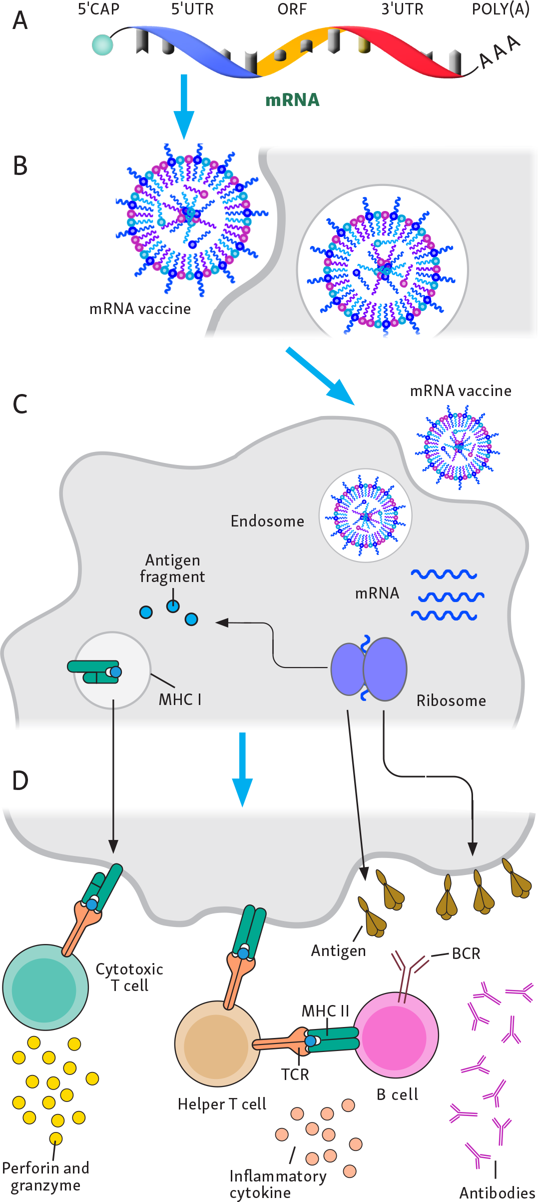
Since these first studies were conducted, several important discoveries have allowed mRNA vaccines to become more popular within the scientific community. These advances are summarized in Figure 1. First, the use of cell-free technology in an in vitro process has made the production of mRNA vaccines more efficient. Another important breakthrough was the incorporation of lipid nanoparticles that surrounded the mRNA molecule in the vaccine, allowing for decreased degradation and enhanced delivery. These lipid nanoparticles are composed of ionizable lipids, improving the safety and extending the circulation time of the mRNA vaccine.4 Following the initial discovery of lipid nanoparticles, new candidate ionizable lipids were examined through large-scale library testing. The result was the discovery of further optimized lipids, such as SM-102 (used in the Moderna COVID-19 mRNA vaccine, or mRNA-1273)5 and ALC-0315 (used in the Pfizer-BioNTech COVID-19 mRNA vaccine, or BNT162b2).6,7 Another important discovery was the identification of the benefits of mRNA modifications such as using modified mRNA nucleosides like pseudouridine. The immune system has evolved pattern recognition receptors (PRRs) that can recognize uridine-rich regions of mRNA. By incorporating pseudouridine into the vaccine, researchers were able to prevent PRR recognition, leading to slower degradation of the mRNA molecule.8 The addition of pseudouridine was first described by Karikó and Weissman and led to the pair receiving the 2023 Nobel Prize in Physiology or Medicine.9-11 Both the Pfizer-BioNTech and Moderna COVID-19 mRNA vaccines included pseudouridine.4 Another major advance in the vaccine field was the use of fusion glycoproteins for respiratory syncytial virus vaccine, which was found to generate improved antibody responses in vaccine recipients.12 All the COVID-19 vaccines (with the exception of the AstraZeneca vaccine) employed a similar fusion-stabilizing mutation in the spike protein that has been demonstrated in preclinical models to improve the induction of neutralizing antibodies.13
Figure 1.
Advances in Messenger RNA Vaccine Technology. Numerous advances have led to the optimized mRNA vaccine technology used today. (A) Improvements in in vitro transcription/cell-free production of mRNA vaccine technology have made vaccine synthesis easier and cost effective. (B) The use of optimized lipid nanoparticles and mRNA modifications, including pseudouridine, has enhanced RNA stability and reduced innate immune breakdown. (C) These advances have resulted in improved uptake of mRNA molecules, leading to ribosomal synthesis of antigen. (D) The ultimate result will be antigen presentation to B cells, leading to antibody responses and antigen fragment presentation to T cells.
Abbreviations: BCR, B-cell receptor; MHC, major histocompatibility complex; mRNA, messenger RNA; TCR, T-cell receptor.
Over time, it was found that mRNA vaccines have advantages over other vaccine platforms. Importantly, mRNA vaccines can be rapidly developed and tailored to new diseases. mRNA vaccines are synthesized using an in vitro transcription process, in which a DNA template is transcribed into mRNA. Once an entity establishes this mRNA vaccine platform, it can easily exchange the open reading frame, or the section of the DNA template that encodes the desired antigen, for a sequence that encodes a new target.14 This strategy can be used to target emerging infectious diseases much more efficiently than other vaccine platforms, resulting in faster vaccine development.14,15 mRNA vaccines are also very immunogenic and have been found to generate robust antibody and CD4+ and CD8+ T-cell responses, as opposed to inactivated or subunit protein vaccines, which will generate responses biased to the humoral immune system.14 Although recombinant virus vaccines can generate strong immune responses, mRNA vaccines may offer improved safety and fewer production challenges.16,17 Also, nucleic acid vaccines, such as mRNA and DNA vaccines, offer improved flexibility in the manufacturing processes, as mentioned previously. However, DNA vaccines require entering the nucleus of a cell to initiate antigen production. Historically, DNA vaccines have been less immunogenic than mRNA vaccines.
Because of the advantages that mRNA vaccines offer, as well as numerous studies showing their safety and immunogenicity in preclinical animal models, researchers began advancing mRNA vaccines to clinical trials in humans. In 2015, one of the first human phase 1 clinical trials targeted the rabies virus.18 Overall, this vaccine generated strong antibody responses with a tolerable safety profile. An mRNA vaccine targeting H10N8 and H7N9 influenza viruses demonstrated antibody seroconversion and tolerability in humans.19 After these successes, groups began preparing to use mRNA vaccine technology but were waiting for the right moment to begin large-scale production.
Vaccine Successes in the COVID-19 Pandemic
In December 2019, health officials began to report an increasing number of pneumonia infections in Wuhan, China.20 As the weeks progressed, it became clear that the new virus causing these infections, later named SARS-CoV-2, posed a substantial health risk. Like its predecessor, the severe acute respiratory syndrome (SARS) virus, the new virus binds to the angiotensin-converting enzyme-2 receptor, but it was ultimately found to be much more transmissible, infecting hundreds of millions and spreading globally. Some of the latest estimates from the World Health Organization indicate that there have been 770 million confirmed infections and approximately 7 million deaths from COVID-19,21 although many experts believe these are likely underestimates given the limitations of identifying cases and reporting these statistics.
Although SARS-CoV-2 had a worldwide impact, the quick development and deployment of vaccines targeting the virus saved countless lives. One model estimates that COVID-19 vaccines saved 14.4 million lives during the second year of the pandemic alone.22 Early collaboration within the scientific community was key to combating this new disease. Such teamwork included the release of the genomic sequence on January 10, 2020, just weeks into the pandemic.23,24 Collaborative relationships were formed as clinical trials testing new therapeutics were started by groups around the world. By the end of 2020, there were numerous medications25-27 and 2 different vaccines28,29 that had been granted emergency use authorization (EUA) by the US Food and Drug Administration (FDA).
Although speed was a priority for the COVID-19 vaccine during a worldwide pandemic, it was important that no shortcuts were taken regarding safety. Numerous decisions and factors led to the rapid development of these vaccines. For example, many phase 1 and phase 2 trials were combined and clinical trialists began designing the phase 3 trial while these earlier trials were ongoing. The researchers also recruited large numbers of study participants and were fortunate that the trials were conducted during periods of relatively high infection rates. Additionally, SARS-CoV-2 proved to be not as formidable a pathogen for vaccine-induced protection as some highly variable viruses such as HIV-1 and hepatitis C.
There are now numerous vaccines targeting SARS-CoV-2. This review focuses primarily on select vaccines that were given monetary support from the US government during the early stages of the pandemic: the Moderna, AstraZeneca, Janssen, Novavax, and Sanofi vaccines. Although the Pfizer-BioNTech vaccine did not receive direct support, the US government agreed to buy it, assuming that it would be efficacious. A summary of these vaccines is shown in the Table. The Pfizer-BioNTech and Moderna vaccines used mRNA vaccine technology.28,29 The AstraZeneca and Janssen vaccines used recombinant adenoviral vector vaccine technology, which involved using a reengineered attenuated virus to deliver SARS-CoV-2 viral DNA that was subsequently translated into proteins and presented to the host immune system.30,31 The Novavax and Sanofi products were protein-based vaccines that included a manufactured version of the SARS-CoV-2 spike protein.32,33 Notably, the Sanofi vaccine did not show efficacy in SARS-CoV-2-naive participants, potentially because of the new SARS-CoV-2 variants emerging. As stated previously, these vaccine trials went through the necessary regulatory processes to ensure patient safety. Importantly, all the vaccines elicited close to 100% protection against severe infection compared with unvaccinated control groups. It should be noted that the original Janssen vaccine trial used only a single dose and the vaccine was initially less protective against severe disease at 85%; however, an additional trial that tested this vaccine with a 2-dose regimen found a level of protection similar to those of the other vaccines tested.34 Long-term data have shown that COVID-19 vaccines are very effective at preventing mortality and severe infection (Figures 2 and 3). On September 22, 2021, the FDA granted additional EUA to the Pfizer-BioNTech, Moderna, and Novavax COVID-19 booster vaccines. As shown in Figure 3, the latest data indicate that vaccinations and these boosters continue to help prevent mortality and severe complications.36
Table.
Overview of SARS-CoV-2 Vaccines
| Vaccine manufacturer | Vaccine type | Dose, regimen | Protection from severe infection, % | Protection from infection, % (95% CI) | Date of approval/EUA |
|---|---|---|---|---|---|
| Pfizer/BioNTech | mRNA | 2 doses, 21 days apart | 100 | 95 (90.3-97.6) | December 11, 2020 |
| Moderna | mRNA | 2 doses, 28 days apart | 100 | 94.1 (89.3-96.8) | December 18, 2020 |
| AstraZeneca | Viral vector | 2 doses, 28 days apart | 100 | 70.4 (54.8-80.6) | December 30, 2020c |
| Janssen | Viral vector | 1 dose | 85b | 66.1 (55.0-74.8)b | February 26, 2021 |
| Novavax | Recombinant protein | 2 doses, 21 days apart | 100 | 89.3 (75.2-95.4) | July 13, 2022 |
| Sanofic | Recombinant protein | 2 doses, 21 days apart | 99 | 64.7 (46.6-77.2) | November 10, 2022d |
a The AstraZeneca vaccine was first approved in Europe and the company never sought EUA from the US Food and Drug Administration.
b The Janssen vaccine was first approved as a single dose on February 27, 2021; a double-dose regimen with improved efficacy was later approved. This approval was withdrawn in May 2023, and Janssen is no longer manufacturing the vaccine.
Although almost all of these COVID-19 vaccines generated a similar degree of protection, there were clear advantages to the mRNA vaccines. The most discussed advantage was the speed and efficiency of the manufacturing process, partly explaining how these vaccines were able to receive EUA from the FDA less than a year after being created. The Pfizer-BioNTech and Moderna mRNA vaccines received this authorization a few months before the Janssen vaccine and a year and a half before the Novavax vaccine. The AstraZeneca vaccine did gain approval only a few weeks after the Pfizer-BioNTech and Moderna vaccines; however, this approval was granted primarily in England and Europe, which have different regulatory processes from those in the US. One example of the difficulties in using other vaccine platforms is evident with the Sanofi vaccine, which was stalled because of a low protein concentration in the first formulation of the vaccine.37 Additionally, the 2 mRNA vaccines appeared to generate mildly improved initial protection from infection compared with the AstraZeneca and Janssen vaccines (see Table).38 It is well documented that protection from infection decreases over time because of a variety of factors, including waning host immune responses and viral div-ersity of SARS-CoV-2. In short, these COVID-19 vaccines elicited a protective immune response against SARS-CoV-2, and the development of these vaccines in less than a year is a testament to the commitment of the scientific community.
Figure 2.
Weekly Confirmed COVID-19 Deaths Per Million People in the US. Weekly confirmed deaths refer to the cumulative number of confirmed deaths over the previous week. Due to varying protocols and challenges in the attribution of the cause of death, the number of confirmed deaths may not accurately represent the true number of deaths caused by COVID-19. Data from the World Health Organization COVID-19 Dashboard, figure adapted with permission from Our World in Data.35
Figure 3.
Weekly COVID-19 Death Rate By Vaccination Status in the US, All Ages. Death rates are calculated as the number of deaths in each group, divided by the total number of people in this group. This is given per 100,000 people. Data from the Centers for Disease Control and Prevention, Vaccine Breakthrough/Surveillance and Analytics Team, figure adapted with permission from Our World in Data.35 Note: The mortality rate for the “all ages” group is age standardized to account for the different vaccination rates of older and younger people.
Difficulties in Creating an HIV-1 Vaccine
In contrast to the SARS-CoV-2 experience, efforts to create an effective vaccine targeting HIV-1 have been unsuccessful, despite decades of research and more than 100 clinical trials. The first HIV-1 vaccine clinical trials performed were aimed at generating antibody responses targeting the surface glycoprotein of HIV-1, known as the HIV-1 envelope (Env).39,40 These early HIV-1 vaccines did induce binding antibody responses, but these antibodies only neutralized a few HIV-1 strains and did not prevent infection in humans exposed to more diverse viral strains, in stark contrast to the COVID-19 vaccines, which induced potent neutralizing antibodies targeting the spike protein of SARS-CoV-2. After these early failures, the strategy changed and the next HIV-1 vaccines targeted intracellular proteins of HIV-1. The hope was that such vaccines would generate CD8+ T-cell responses that might not prevent infection but could protect against HIV-1 progression and AIDS.41,42 Although such findings were demonstrated in preclinical nonhuman primate models,43 the human efficacy study that tested this concept failed to demonstrate protection against infection or an impact on plasma HIV RNA level in those infected. After these clinical trials, the next study conducted was the RV144 (Thai Phase 3 clinical trial), which showed modest efficacy (31.2% at 42 months) after statistical adjustments and may represent the most successful HIV-1 vaccine trial to date.44 This vaccine generated both antibody and CD4+ T-cell responses toward HIV-1, and it was later found that increased levels of protection correlated with antibodies specific for a region of Env known as the V1V2 loop.44 Despite this promising result, a more recent trial performed in South Africa known as HVTN (HIV Vaccine Trials Network) 702 was intended to build on these results using a vaccine strategy similar to that of RV144, but ultimately no efficacy was demonstrated.45
There are numerous reasons why researchers have had such difficulties with creating an effective HIV-1 vaccine. HIV-1 vaccines generate antibody responses, but, in contrast to other viral vaccines such as those targeting COVID-19, the antibodies are poorly neutralizing and do not prevent HIV-1 infection. This phenomenon is due at least in part to the remarkable viral diversity of HIV-1.46,47 As these mutations arise over the course of chronic infection within a host, specific strains will have the ability to undergo immune escape and evade the host immune response. Supporting this theory is that numerous HIV-1 mutations have been proven to represent escape from antibody and CD8+ T-cell responses.48,49 Our group has also shown that HIV-1 can mutate, or undergo adaptation, in response to CD4+ T-cell responses,50 and that these HIV-1 adaptations to CD4+ and CD8+ T-cell responses can affect HIV-1 vaccine responses.51,52 The ability of HIV-1 to mutate quickly is also the reason the virus develops resistance to certain drugs, resulting in the clinical treatment of HIV-1 with a cocktail of 3 antiretroviral medications.53 An additional hurdle is that after infection, HIV-1 integrates into the host genome and causes latent infection. As a result, to ultimately be effective, an HIV-1 vaccine will likely need to completely prevent infection, as opposed to preventing only symptomatic infection or severe infection, as with vaccines for other viruses. As a result, the task of creating an effective vaccine for HIV-1 poses one of the greatest challenges vaccine researchers have faced.
However, there is reason for hope. Two recent studies, collectively referred to as the AMP (antibody-mediated protection) study, investigated whether protection from HIV-1 infection could be achieved via passive immunization of an HIV-specific antibody.54 Unlike traditional vaccination strategies that rely on the host immune system to produce antibodies, recipients in this study were passively immunized with an antibody targeting Env. This antibody was the broadly neutralizing antibody (bNAb) called VRC01, which has been shown to target numerous strains of HIV-1.54 Unfortunately, no overall protection against infection following bNAb injection was demonstrated. However, analyses of the results showed that participants receiving this antibody were less likely to be infected with VRC01-sensitive HIV-1 strains, suggesting that the bNAb was providing some level of protection against certain strains of HIV-1. This is an important finding and suggests that for future HIV-1 vaccines to be effective, the immune response would likely need to generate multiple bNAbs with complementary mechanisms of action, similar to what is achieved with combination antiretroviral therapy for HIV-1 treatment. Follow-up studies are being discussed that would confirm this hypothesis by investigating whether passive immunization with multiple bNAbs could prevent HIV-1 infection.
Promising New HIV-1 Vaccine Strategies and the Potential Role of mRNA Vaccine Technology
Although we now have a better idea of what may be needed, the task of creating an HIV-1 vaccine remains daunting. bNAbs are typically produced in only a minority of individuals after years of chronic HIV-1 infection. To generate HIV-specific bNAbs, a vaccine would need to mimic the process of what happens over years of HIV-1 infection. This approach has led to one of the most promising strategies, which is to create bNAbs by sequential immunization, involving a vaccine with 3 distinct steps to mirror the development of bNAb-producing B cells (Figure 4). The first step is called “priming,” which involves germline targeting and expanding the first B-cell precursors. Although these B cells do not have neutralizing antibody capacity, they do have the potential to produce HIV-1 bNAbs if subsequently boosted with the correct antigen. This boosting, or second step, will involve “shepherding” these precursors through B-cell development, and the final step, termed “polishing,” will mature these cells into bNAb-producing plasma cells. Recent studies have been successful in priming naive B cells in order to expand B-cell precursors with the potential to specifically produce VRC01 bNAbs.55 Ongoing research is focused on using this strategy or a similar framework to expand other B-cell precursors capable of targeting other regions of Env. Although many bNAbs target the CD4 binding site of HIV-1 Env, several other bNAb targets have been identified, including V2 apex, V3 glycan, fusion peptide, and the membrane-proximal external region. Recent reviews have discussed these findings in detail.56 Ultimately, based on the findings from the AMP study, an HIV-1 vaccine may be able to elicit protection if it can generate bNAbs that target several complementary HIV-1 Env sites.
Figure 4.
Strategies for Future HIV-1 Vaccine Trials. A successful HIV-1 vaccine will likely need to use a multicomponent approach. (A) The most promising strategy to date involves sequential immunization, which uses a 3-step approach of priming/germline targeting, then shepherding, and finally polishing to generate bNAb-secreting plasma cells. (B) In addition, the HIV vaccine would involve dendritic cell presentation of antigen to both CD4+ and CD8+ T cells. Optimal CD4+ T cells would form robust T-follicular helper-cell responses that assist in shepherding and polishing the B-cell response. (C) Optimal CD8+ T-cell responses, potentially using human leukocyte antigen (HLA)-E-specific responses, would then be able to assist in killing any virally infected cell. Abbreviations: GC, germinal center; bNAb, broadly neutralizing antibody.
Although B-cell and antibody generation has been a recent focus in the HIV-1 vaccine field, optimization of T-cell responses may also play an important role in HIV-1 vaccine design. A specific subset of CD4+ T cells known as T follicular helper (Tfh) cells are found in the germinal centers of lymph nodes and may be crucial to maturation of B-cell precursors into bNAb-producing plasma cells. Supporting this idea is that the RV144 trial indicated a correlation between polyfunctional Env-specific CD4+ T cells and decreased risk of infection.57 More recent studies have shown that induction of strong CD4+ Tfh cell response was required to induce bNAbs.58,59 Future research should investigate adjuvants and other vaccine strategies capable of stimulating Tfh-dominant responses.
Additionally, it may be possible to improve HIV-1 vaccine responses by harnessing CD8+ T cells. Although previous HIV-1 vaccines aimed at generating CD8+ T-cell responses were shown to be ineffective at providing protection from infection, there is evidence to suggest that CD8+ T cells can play a role in HIV-1 vaccines. In HVTN 505, a previous HIV-1 vaccine efficacy trial, CD8+ T-cell responses targeting Env correlated with decreased risk of infection.60 Also, there has been promise in investigating HLA-E-specific CD8+ T-cell responses. These responses were first described with a cytomegalovirus viral vector vaccine that induced CD8+ T cells restricted by the HLA-E analogue in simian immunodeficiency virus animal models.61 Such responses were shown to be essential to protect against simian immunodeficiency virus infection.62 These preclinical animal vaccine trials are encouraging, and human clinical studies are currently ongoing. However, the cytomegalovirus viral vector will be a live attenuated vaccine, with greater challenges in manufacturing and potentially increased adverse effects.
Although many HIV-1 vaccine studies have used other vaccine types, mRNA vaccine technology can play a crucial role in the ongoing search for an effective HIV-1 vaccine. Many experts believe that mRNA vaccines are optimal for testing new vaccine strategies because they can deliver complex multipart immunogens. An effective HIV-1 vaccine will likely need to generate complementary bNAbs while also stimulating Tfh and CD8+ T-cell responses. This broad approach will require investigation of complementary strategies. mRNA vaccines provide a good platform because they generate strong T-cell and antibody responses. Because various mRNA vaccines can be created quickly, these new strategies could be investigated more efficiently using the mRNA platform, providing the field with answers regarding how to optimize the next generation of vaccines. mRNA vaccines may also produce an improved immune response compared with other vaccine platforms. For instance, previous studies have shown that lipid nanoparticle-enclosed mRNA can induce potent Tfh responses.63·64 Ongoing studies are investigating whether Env trimer nanoparticle multimers can be formed using the mRNA platform.65,66 Inclusion of the transmembrane domain of HIV-1 Env in mRNA vaccines could lead to the generation of a membrane-bound Env that may prove to be beneficial, as it will lead to presentation to the immune system in its more natural form. Future studies should also investigate whether mRNA vaccines can generate HLA-E CD8+ T-cell responses by targeting dendritic cells, as these antigen-presenting cells have increased expression of HLA-E. In summary, mRNA vaccines can quickly test new strategies and have the potential to generate a multifaceted, complex immune response that will ultimately be required to protect against HIV-1 infection.
Future Advances in mRNA Vaccine Technology
In addition to HIV-1, mRNA vaccine technology is being investigated for the prevention of other infections, with ongoing clinical trials examining mRNA vaccines targeting respiratory syncytial virus, influenza viruses, Zika virus, rabies virus, Ebola virus, and malaria.4 Recent advances in mRNA vaccine technology may prove to be beneficial for preventing these infections as well.
One of these advances is self-amplifying mRNA. This strategy involves including a viral replicase gene in the vaccine open reading frame in addition to a designed antigen target.67 In a study performed in mice, this strategy led to a 10-fold increase in protein expression and increased the duration of antigen detection from 2 days to 10 days.68 Using this strategy not only increases immunogenicity by prolonging antigen presentation but also decreases the amount of mRNA needed. This would decrease PRR recognition and the innate immune response, leading to stronger adaptive immune responses and ultimately improved antibody responses, such as generation of bNAbs by an HIV-1 vaccine. This strategy may also induce longer-lasting antibodies, which has been a particular problem with the existing mRNA platforms.69
Other ongoing research is focused on optimizing mRNA vaccines to target specific tissues and cells. Achieving this would allow researchers to target immune response toward the area where infection is most likely to occur. For SARS-CoV-2 and influenza viruses, vaccine immune responses in the upper respiratory system are crucial, whereas genital and rectal mucosal immune responses are much more important in combating HIV-1. Another strategy involves using mRNA to target specific immune cells. As mentioned previously, specific lipid nanoparticle formulations have been found to stimulate stronger Tfh-type responses.63 Other groups are investigating how to generate a strong dendritic cell response, which can lead to improved antigen presentation and stronger overall immune responses.
Several limitations to mRNA vaccine technology merit discussion. One is temperature storage requirements, which currently are temperatures of-20 °C or colder. This will be a major obstacle for HIV-1 vaccines, as much of the developing world where HIV-1 is most prevalent does not have the infrastructure required to store vaccines at this temperature. From an immunologic standpoint, there is also the limitation that antibody responses generated from mRNA vaccines alone appear to be less durable compared with vaccination in the context of prior infection.70 Although continuing to boost vaccine responses is possible, this may not be a cost-effective method when trying to vaccinate a large number of individuals. It is also important to note that the mRNA technology is still relatively new and only COVID-19 vaccines are FDA approved using this platform. Time will tell whether the mRNA platform can be consistently used to develop vaccines targeting other pathogens.
Conclusion
The use of mRNA vaccine technology to create safe and effective vaccines quickly during the COVID-19 pandemic has been one of the most remarkable achievements of medical research of our generation. Meanwhile, HIV-1 vaccine efforts fail to elicit effective protection. However, the HIV-1 vaccine field now has a clear goal to create a vaccine that induces bNAbs, and there are several new strategies that show promise in this regard. It is likely that a multifaceted immune response will be needed, generating potent HIV-specific bNAbs, an optimal CD4+ T-cell response, and a strong CD8+ T-cell response. mRNA vaccine technology is a powerful vaccine platform to test these new strategies, with the potential to benefit ongoing HIV-1 vaccine research efforts.
Notes
This article was based on a presentation by Paul A. Goepfert, MD, at the IAS-USA Annual Update on HIV Management in Atlanta, Georgia on March 31, 2023.
The IAS-USA will identify and resolve ahead of time any possible conflicts of interest that may influence continuing medical education (CME) activities with regard to exposition or conclusion. All financial relationships with ineligible companies for the authors and reviewers are listed below.
Financial relationships with ineligible companies in the past 24 months: Dr Files reported no relevant financial relationships with ineligible companies. (Updated April 4, 2024) Dr Goepfert reported no relevant financial relationships with ineligible companies. (Updated April 4, 2024)
References
S Brenner, F Jacob, M Meselson. An unstable intermediate carrying information from genes to ribosomes for protein synthesis. Nature. 1961; 190: 576–581. doi:10.1038/190576a0
F Martinon, S Krishnan, G Lenzen, et al. Induction of virus-specific cytotoxic T lymphocytes in vivo by liposome-entrapped mRNA. Eur J Immunol. 1993; 23(7): 1719–1722. doi:10.1002/eji.1830230749
N Chaudhary, D Weissman, KA Whitehead. mRNA vaccines for infectious diseases: principles, delivery and clinical translation. Nat Rev Drug Discov. 2021; 20(11): 817–838. doi:10.1038/s41573-021-00283-5
S Sabnis, ES Kumarasinghe, T Salerno, et al. A novel amino lipid series for mRNA delivery: improved endosomal escape and sustained pharmacology and safety in non-human primates. Mol Ther. 2018; 26(6): 1509–1519. doi:10.1016/j.ymthe.2018.03.010
MJ Hope, B Mui, PJC Lin, et al, inventors; Acuitas Therapeutics Inc, assignee. Lipid nanoparticle formulations. International patent WO-2018081480-A1. May 3, 2018. Accessed February 22, 2024. https://pubchem.ncbi.nlm.nih.gov/patent/WO-2018081480-A1#section=Abstract
MD Buschmann, MJ Carrasco, S Alishetty, M Paige, MG Alameh, D Weissman. Nanomaterial delivery systems for mRNA vaccines. Vaccines (Basel). 2021; 9(1): 65. doi:10.3390/vaccines9010065
KA Hajj, KA Whitehead. Tools for translation: non-viral materials for therapeutic mRNA delivery. Nat Rev Mat. 2017; 2: 17056. doi:10.1038/natrevmats.2017.56
K Karikó, M Buckstein, H Ni, D Weissman. Suppression of RNA recognition by Toll-like receptors: the impact of nucleoside modification and the evolutionary origin of RNA. Immunity. 2005; 23(2): 165–175. doi:10.1016/j.immuni.2005.06.008
K Karikó, H Muramatsu, FA Welsh, et al. Incorporation of pseudouridine into mRNA yields superior nonimmunogenic vector with increased translational capacity and biological stability. Mol Ther. 2008; 16(11): 1833–1840. doi:10.1038/mt.2008.200
BR Anderson, H Muramatsu, SR Nallagatla, et al. Incorporation of pseudouridine into mRNA enhances translation by diminishing PKR activation. Nucleic Acids Res. 2010; 38(17): 5884–5892. doi:10.1093/nar/gkq347
JS McLellan, M Chen, MG Joyce, et al. Structure-based design of a fusion glycoprotein vaccine for respiratory syncytial virus. Science. 2013; 342(6158): 592–598. doi:10.1126/science.1243283
KS Corbett, DK Edwards, SR Leist, et al. SARS-CoV-2 mRNA vaccine design enabled by prototype pathogen preparedness. Nature. 2020; 586(7830): 567–571. doi:10.1038/s41586-020-2622-014.
S Son, K Lee. Development of mRNA vaccines/therapeutics and their delivery system. Mol Cells. 2023; 46(1): 41–47. doi:10.14348/molcells.2023.2165
R Verbeke, I Lentacker, SC De Smedt, H Dewitte. Three decades of messenger RNA vaccine development. Nano Today. 2019; 28: 100766. doi:10.1016/j.nantod.2019.100766
A Buoninfante, A Andeweg, AT Baker, et al. Understanding thrombosis with thrombocytopenia syndrome after COVID-19 vaccination. NPJ Vaccines. 2022; 7(1): 141. doi:10.1038/s41541-022-00569-8
ML Lee, JMP Bautista. Guillain-Barré syndrome following the administration of adenovirus vector-based COVID-19 vaccine. Cureus. 2023; 15(7): e42316. doi:10.7759/cureus.42316
M Alberer, U Gnad-Vogt, HS Hong, et al. Safety and immunogenicity of a mRNA rabies vaccine in healthy adults: an open-label, non-randomised, prospective, first-in-human phase 1 clinical trial. Lancet. 2017; 390(10101): 1511–1520. doi:10.1016/S0140-6736(17)31665-3
K Bahl, JJ Senn, O Yuzhakov, et al. Preclinical and clinical demonstration of immunogenicity by mRNA vaccines against H10N8 and H7N9 influenza viruses. Mol Ther. 2017; 25(6): 1316–1327. doi:10.1016/j.ymthe.2017.03.035
World Health Organization. WHO statement regarding cluster of pneumonia cases in Wuhan, China. January 9, 2020. Accessed February 22, 2024. https://www.who.int/hongkongchina/news/detail/09-01-2020-who-statement-regarding-cluster-of-pneumonia-cases-in-wuhan-china
World Health Organization. WHO coronavirus (COVID-19) dashboard. September 24, 2023. Accessed February 22, 2024. https://covid19.who.int/
OJ Watson, G Barnsley, J Toor, AB Hogan, P Winskill, AC Ghani. Global impact of the first year of COVID-19 vaccination: a mathematical modelling study. Lancet Infect Dis. 2022; 22(9): 1293–1302. doi:10.1016/S1473-3099(22)00320-6
EC Holmes. Re: Zhang YZ. Initial genome release of novel coronavirus. January 10, 2020. Accessed February 22, 2024. https://virological.org/t/novel-2019-coronavirus-genome/319
X Xu, P Chen, J Wang, et al. Evolution of the novel coronavirus from the ongoing Wuhan outbreak and modeling of its spike protein for risk of human transmission. Sci China Life Sci. 2020; 63(3): 457–460. doi:10.1007/s11427-020-1637-5
JH Beigel, KM Tomashek, LE Dodd, et al. Remdesivir for the treatment of COVID-19—final report. N Engl J Med. 2020; 383(19): 1813–1826. doi:10.1056/NEJMoa2007764
AC Kalil, TF Patterson, AK Mehta, et al. Baricitinib plus remdesivir for hospitalized adults with COVID-19. N Engl J Med. 2021; 384(9): 795–807. doi:10.1056/NEJMoa2031994
DM Weinreich, S Sivapalasingam, T Norton, et al. REGEN-COV antibody combination and outcomes in outpatients with COVID-19. N Engl J Med. 2021; 385(23): e81. doi:10.1056/NEJMoa2108163
LR Baden, HM El Sahly, B Essink, et al. Efficacy and safety of the mRNA-1273 SARS-CoV-2 vaccine. N Engl J Med. 2021; 384(5): 403–416. doi:10.1056/NEJMoa2035389
FP Polack, SJ Thomas, N Kitchin, et al. Safety and efficacy of the BNT162b2 mRNA COVID-19 vaccine. N Engl J Med. 2020; 383(27): 2603–2615. doi:10.1056/NEJMoa2034577
J Sadoff, G Gray, A Vandebosch, et al. Safety and efficacy of single-dose Ad26.COV2.S vaccine against COVID-19. N Engl J Med. 2021; 384(23): 2187–2201. doi:10.1056/NEJM-oa2101544
M Voysey, SAC Clemens, SA Madhi, et al. Safety and efficacy of the ChAdOx1 nCoV-19 vaccine (AZD1222) against SARS-CoV-2: an interim analysis of four randomised controlled trials in Brazil, South Africa, and the UK. Lancet. 2021; 397(10269): 99–111. doi:10.1016/S0140-6736(20)-32661-1
PT Heath, EP Galiza, DN Baxter, et al. Safety and efficacy of NVX-CoV2373 COVID-19 vaccine. N Engl J Med. 2021; 385(13): 1172–1183. doi:10.1056/NEJMoa2107659
GH Dayan, N Rouphael, SR Walsh, et al. Efficacy of a bivalent (D614 + B.1.351) SARS-CoV-2 recombinant protein vaccine with AS03 adjuvant in adults: a phase 3, parallel, randomised, modified double-blind, placebo-controlled trial. Lancet Respir Med. 2023; 11(11): 975–990. doi:10.1016/S2213-2600(23)00263-1
K Hardt, A Vendebosch, J Sadoff, et al. Efficacy, safety, and immunogenicity of a booster regimen of Ad26.COV2.S vaccine against COVID-19 (eNSEMBLE2): results of a randomised, double-blind, placebo-controlled, phase 3 trial. Lancet Infect Dis. 2022; 22(12): 1703–1715. doi:10.1016/S1473-3099(22)00506-0
E Mathieu, H Ritchie, L Rodés-Guirao, et al. Coronavirus pandemic (COVID-19). Our World in Data website. Accessed February 22, 2024. https://ourworldindata.org/coronavirus
Centers for Disease Control and Prevention. Monthly age-adjusted rates of COVID-19-associated hospitalization by vaccination status. March 15, 2023. Accessed February 22, 2024. https://www.cdc.gov/coronavirus/2019-ncov/covid-data/covid-net/hospitalizations-by-vaccination-status-report.pdf
PA Goepfert, B Fu, AL Chabanon, et al. Safety and immunogenicity of SARS-CoV-2 recombinant protein vaccine formulations in healthy adults: interim results of a randomised, placebo-controlled, phase 1-2, dose-ranging study. Lancet Infect Dis. 2021; 21(9): 1257–1270. doi:10.1016/S1473-3099(21)00147-X
MG Thompson, JL Burgess, AL Naleway, et al. Prevention and attenuation of COVID-19 with the BNT162b2 and mRNA-1273 vaccines. N Engl J Med. 2021; 385(4): 320–329. doi:10.1056/NEJMoa2107058
NM Flynn, DN Forthal, CD Harro, et al. Placebo-controlled phase 3 trial of a recombinant glycoprotein 120 vaccine to prevent HIV-1 infection. J Infect Dis. 2005; 191(5): 654–665. doi:10.1086/428404
P Pitisuttithum, P Gilbert, M Gurwith, et al. Randomized double-blind, placebo-controlled efficacy trial of a bivalent recombinant glycoprotein 120 HIV-1 vaccine among injection drug users in Bangkok, Thailand. J Infect Dis. 2006; 194(12): 1661–1671. doi:10.1086/508748
SP Buchbinder, DV Mehrotra, A Duerr, et al. Efficacy assessment of a cell-mediated immunity HIV-1 vaccine (the Step study): a double-blind, randomised, placebo-controlled, test-of-concept trial. Lancet. 2008; 372(9653): 1881–1893. doi:10.1016/S0140-6736(08)61591-3
A Duerr, Y Huang, S Buchbinder, et al. Extended follow-up confirms early vaccine-enhanced risk of HIV acquisition and demonstrates waning effect over time among participants in a randomized trial of recombinant adenovirus HIV vaccine (Step study). J Infect Dis. 2012; 206(2): 258–266. doi:10.1093/infdis/jis342
JW Shiver, TM Fu, L Chen, et al. Replication-incompetent adenoviral vaccine vector elicits effective anti-immunodeficiency-virus immunity. Nature. 2002; 415(6869): 331–335. doi:10.1038/415331a
S Rerks-Ngarm, P Pitisuttithum, S Nitayaphan, et al. Vaccination with ALVAC and AIDSVAX to prevent HIV-1 infection in Thailand. N Engl J Med. 2009; 361(23): 2209–2220. doi:10.1056/NEJMoa0908492
GE Gray, LG Bekker, F Laher, et al. Vaccine efficacy of ALVAC-HIV and bivalent subtype C gp120-MF59 in adults. N Engl J Med. 2021; 384(12): 1089–1100. doi:10.1056/NEJM-oa2031499
JW Rausch, AA Capoferri, MG Katusiime, SC Patro, MF Kearney. Low genetic diversity may be an Achilles heel of SARS-CoV-2. Proc Natl Acad Sci U S A. 2020; 117(40): 24614–24616. doi:10.1073/pnas.2017726117
W Fischer, EE Giorgi, S Chakraborty, et al. HIV-1 and SARS-CoV-2: patterns in the evolution of two pandemic pathogens. Cell Host Microbe. 2021; 29(7): 1093–1110. doi:10.1016/j.chom.2021.05.012
P Borrow, H Lewicki, X Wei, et al. Antiviral pressure exerted by HIV-1-specific cytotoxic T lymphocytes (CTLs) during primary infection demonstrated by rapid selection of CTL escape virus. Nat Med. 1997; 3(2): 205–211. doi:10.1038/nm0297-205
X Wei, JM Decker, S Wang, et al. Antibody neutralization and escape by HIV-1. Nature. 2003; 422(6929): 307–312. doi:10.1038/nature01470
N Erdmann, VY Du, J Carlson, et al. HLA dass-II associated HIV polymorphisms predict escape from CD4+ T cell responses. PLoS Pathog. 2015; 11(8): e1005111. doi:10.1371/journal.ppat.1005111
S Boppana, S Sterrett, J Files, et al. HLA-I associated adaptation dampens CD8 T-cell responses in HIV Ad5-vectored vaccine recipients. J Infect Dis. 2019; 220(10): 1620–1628. doi:10.1093/infdis/jiz368
JK Files, S Sterrett, S Henostroza, et al. HLA-II-associated HIV-1 adaptation decreases cD4+ T-cell responses in HIV-1 vaccine recipients. J Virol. 2022; 96(17): e0119122. doi:10.1128/jvi.01191-22
L Corey, PB Gilbert, M Juraska, et al. Two randomized trials of neutralizing antibodies to prevent HIV-1 acquisition. N Engl J Med. 2021; 384(11): 1003–1014. doi:10.1056/NEJMoa2031738
DJ Leggat, KW Cohen, JR Willis, et al. Vaccination induces HIV broadly neutralizing antibody precursors in humans. Science. 2022; 378(6623): eadd6502. doi:10.1126/science.add6502
BF Haynes, K Wiehe, P Borrow, et al. Strategies for HIV-1 vaccines that induce broadly neutralizing antibodies. Nat Rev Immunol. 2023; 23(3): 142–158. doi:10.1038/s41577-022-00753-w
L Lin, G Finak, K Ushey, et al. COMPASS identifies T-cell subsets correlated with clinical outcomes. Nat Biotechnol. 2015; 33(6): 610–616. doi:10.1038/nbt.3187
M Locci, C Havenar-Daughton, E Landais, et al. Human circulating PD-1+CXCR3-CXCR5+ memory Tfh cells are highly functional and correlate with broadly neutralizing HIV antibody responses. Immunity. 2013; 39(4): 758–769. doi:10.1016/j.immuni.2013.08.031
MA Moody, I Pedroza-Pacheco, NA Vandergrift, et al. Immune perturbations in HIV-1-infected individuals who make broadly neutralizing antibodies. Sci Immunol. 2016; 1(1): aag0851. doi:10.1126/sciimmunol.aag0851
HE Janes, KW Cohen, N Frahm, et al. Higher T-cell responses induced by DNA/rAd5 HIV-1 preventive vaccine are associated with lower HIV-1 infection risk in an efficacy trial. J Infect Dis. 2017; 215(9): 1376–1385. doi:10.1093/infdis/jix086
SG Hansen, JB Sacha, CM Hughes, et al. Cytomegalovirus vectors violate CD8+ T cell epitope recognition paradigms. Science. 2013; 340(6135): 1237874. doi:10.1126/science.1237874
SG Hansen, MH Hancock, D Malouli, et al. Myeloid cell tropism enables MHC-E-restricted CD8+ T cell priming and vaccine efficacy by the RhCMV/SIV vaccine. Sci Immunol. 2022; 7(72): eabn9301. doi:10.1126/sciimmunol.abn9301
N Pardi, MJ Hogan, MS Naradikian, et al. Nucleoside-modified mRNA vaccines induce potent T follicular helper and germinal center B cell responses. J Exp Med. 2018; 215(6): 1571–1588. doi:10.1084/jem.20171450
MG Alameh, I Tombácz, E Bettini, et al. Lipid nanoparticles enhance the efficacy of mRNA and protein subunit vaccines by inducing robust T follicular helper cell and humoral responses. Immunity. 2021; 54(12): 2877–2892.e7. doi:10.1016/j.immuni.2021.11.001
Z Mu, K Wiehe, KO Saunders, et al. mRNA-encoded HIV-1 Env trimer ferritin nanoparticles induce monoclonal antibodies that neutralize heterologous HIV-1 isolates in mice. Cell Rep. 2022; 38(11): 110514. doi:10.1016/j.celrep.-2022.110514
Z Mu, BF Haynes, DW Cain. HIV mRNA vaccines-progress and future paths. Vaccines (Basel). 2021; 9(2): 134. doi:10.3390/vaccines9020134
A Cagigi, K Loré. Immune responses induced by mRNA vaccination in mice, monkeys and humans. Vaccines (Basel). 2021; 9(1): 61. doi:10.3390/vaccines9010061
FW Johanning, RM Conry, AF LoBuglio, et al. A Sindbis virus mRNA polynucleotide vector achieves prolonged and high level heterologous gene expression in vivo. Nucleic Acids Res. 1995; 23(9): 1495–1501. doi:10.1093/nar/23.9.1495
DR Feikin, MM Higdon, LJ Abu-Raddad, et al. Duration of effectiveness of vaccines against SaRS-CoV-2 infection and COVID-19 disease: results of a systematic review and meta-regression. Lancet. 2022; 399(10328): 924–944. doi:10.1016/S0140-6736(22)00152-0
D Zhong, S Xiao, AK Debes, et al. Durability of antibody levels after vaccination with mRNA SARS-CoV-2 vaccine in individuals with or without prior infection. JAMA. 2021; 326(24): 2524–2526. doi:10.1001/jama.2021.19996