Introduction
Despite major advances in HIV treatment coverage, more than 4 million people globally have advanced HIV disease, and more than 600,000 die each year.1,2 Opportunistic infections continue to be a substantial cause of morbidity and mortality among these individuals, accounting for the majority of these deaths.1,2 This problem is not limited to only low- and middle-income countries; more than 1 in 5 people diagnosed with HIV in the US are diagnosed with late-stage disease, and 24% of people diagnosed with HIV in the US are not retained in care.3 Furthermore, although hospitalizations from opportunistic infections in the US have decreased substantially, the proportion of hospitalizations that results in mortality has remained stable.4 Among opportunistic infections, tuberculosis and cryptococcal meningitis remain the leading causes of death, accounting for 40% and 19% of deaths worldwide among adults with HIV, respectively.1,5,6
This compendium of images outlines the presentation, diagnosis, and management of study participants who were evaluated under 2 clinical protocols (NCT00286767 and NCT02147405) from 2004 until 2024. These protocols enrolled people with advanced HIV (CD4+ count <100 cells/μL before initiating antiretroviral therapy [ART]) or who had suspected immune reconstitution inflammatory syndrome (IRIS). The case summaries are accompanied by clinical photography, endoscopic images, retinal photography, radiographic imaging, microbiologic images, and pathologic images to illustrate the patients’ clinical courses.
Bartonellosis
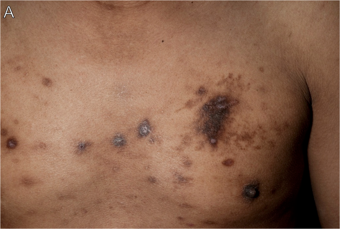
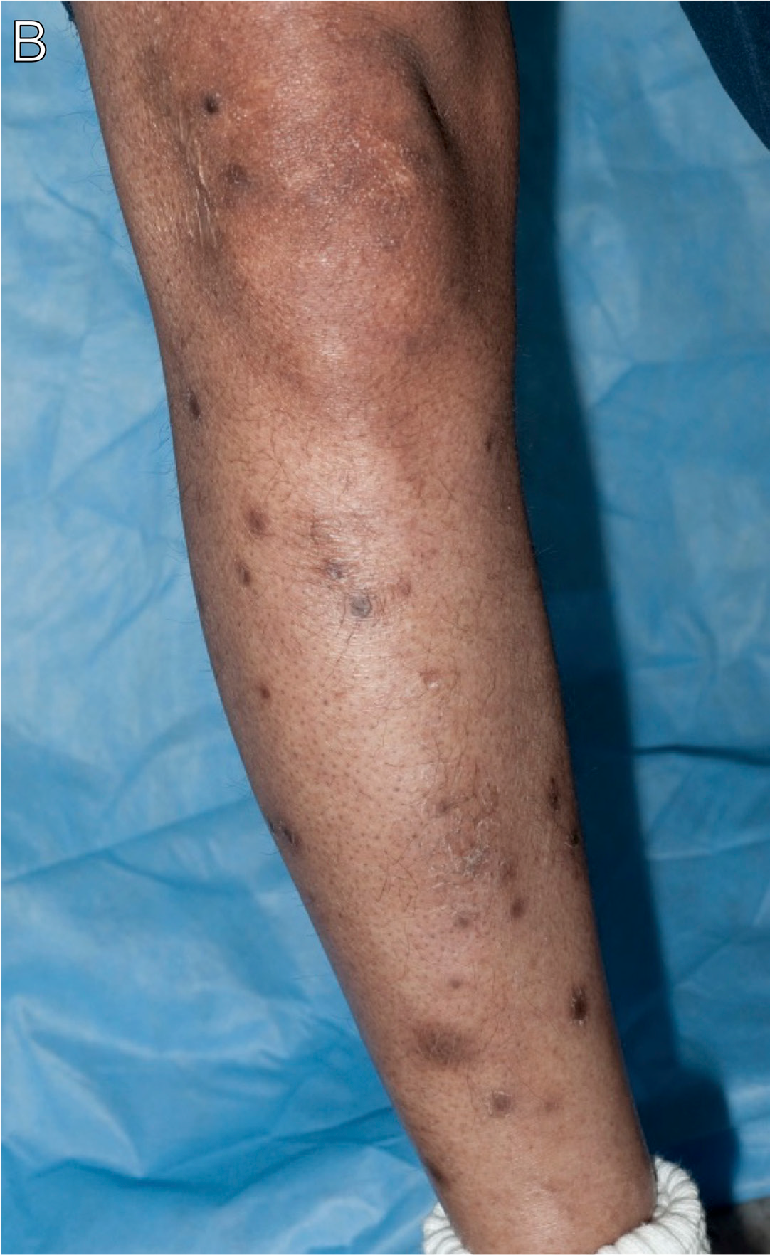
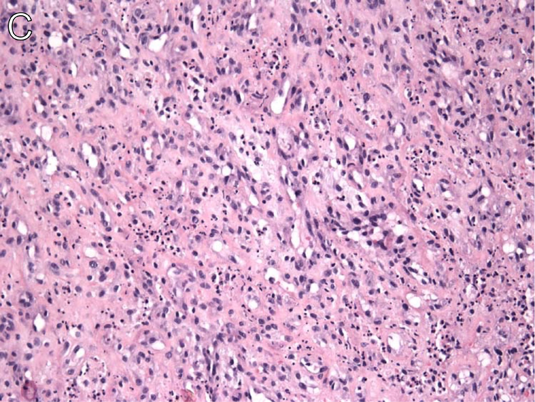
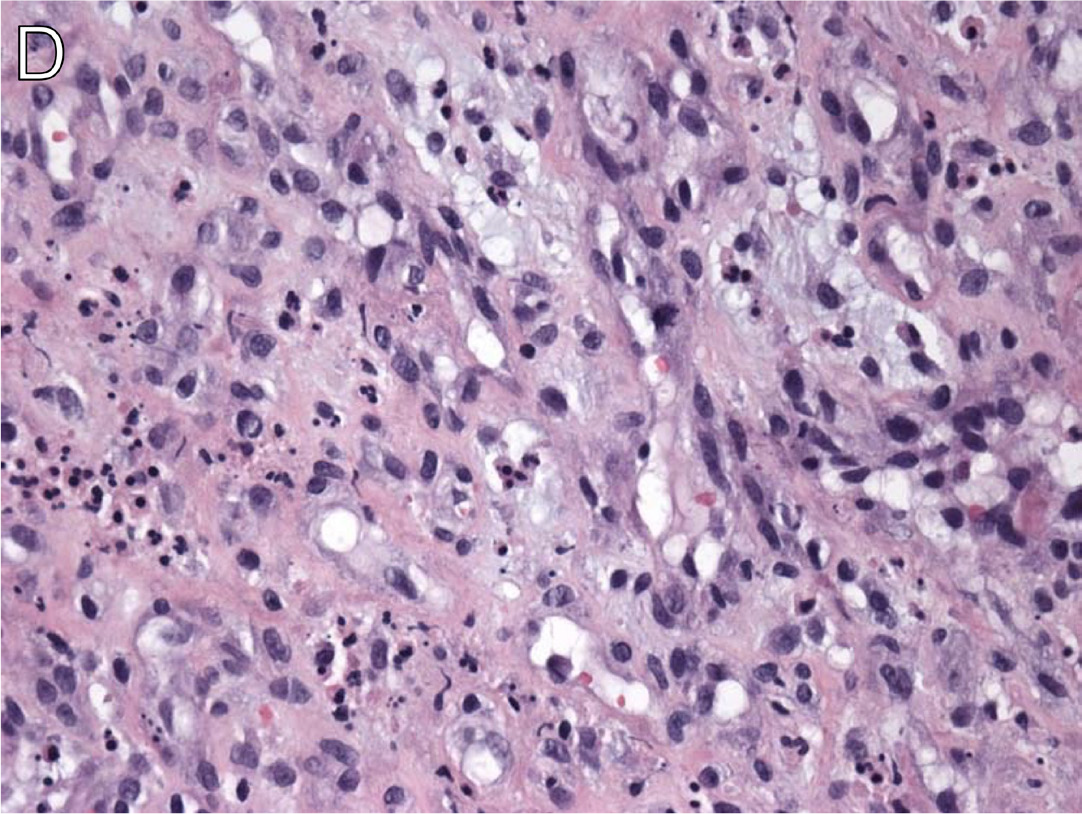
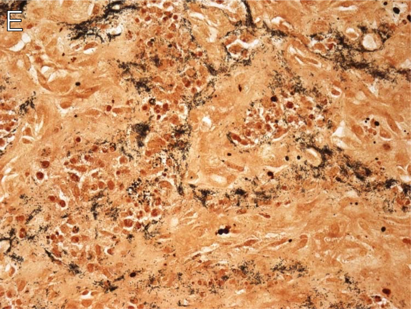
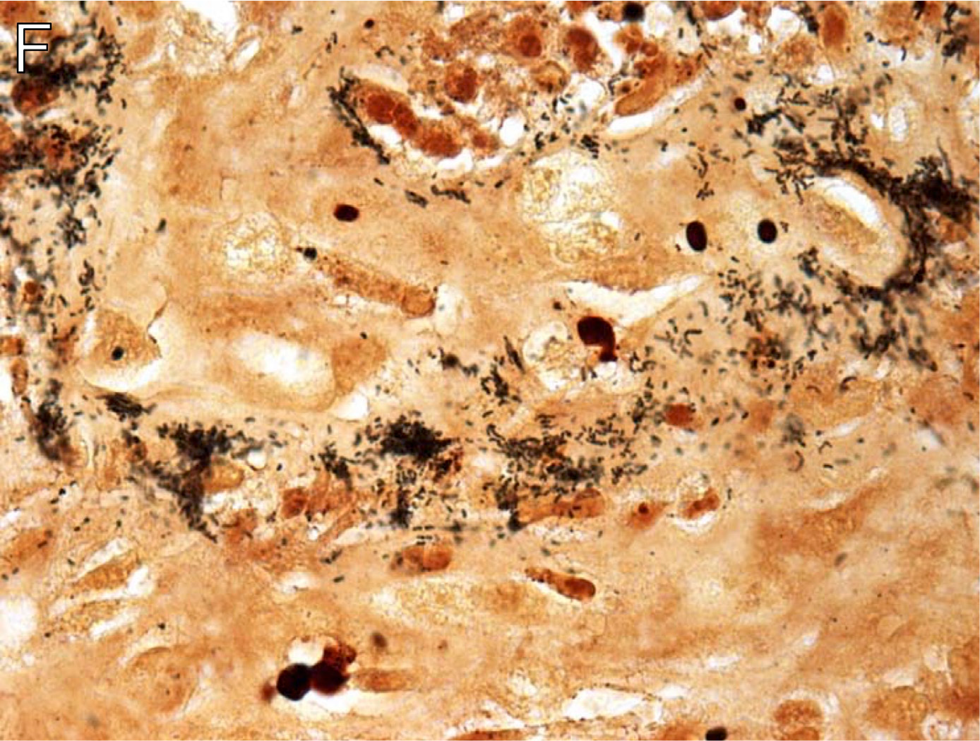
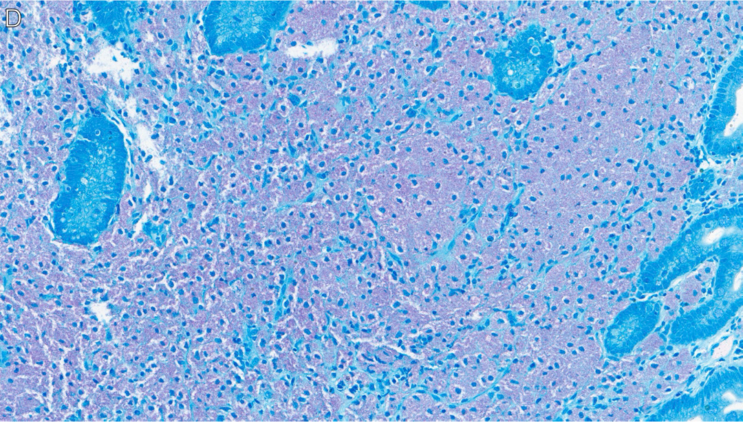
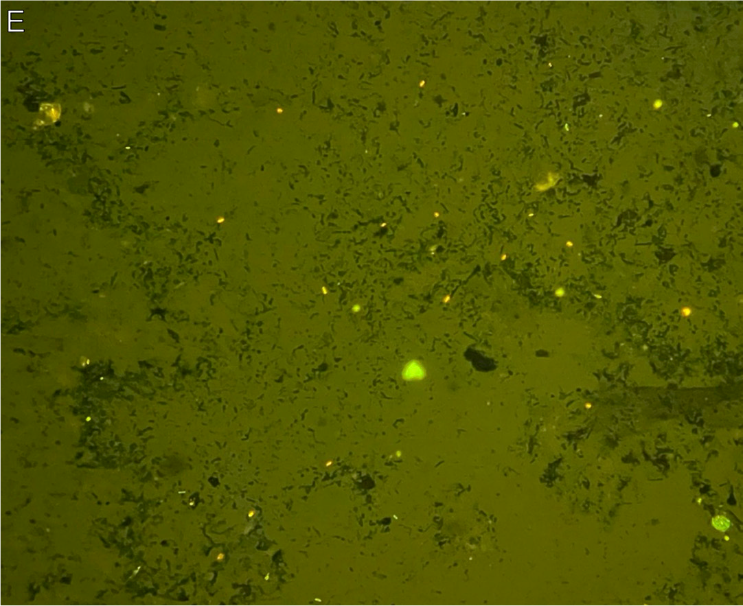
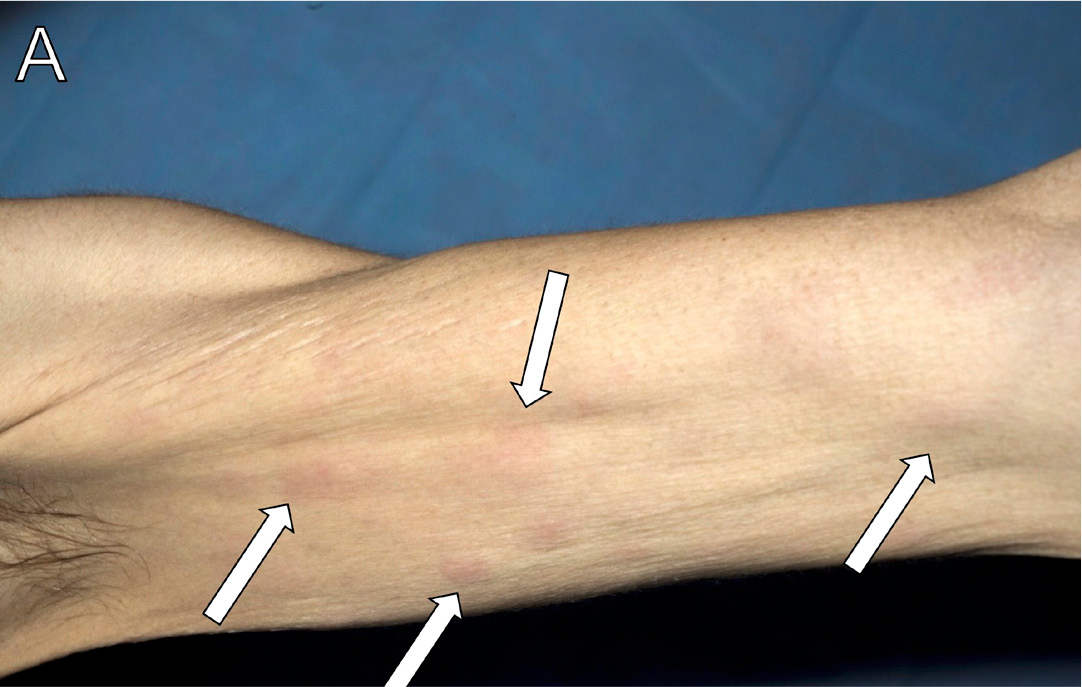
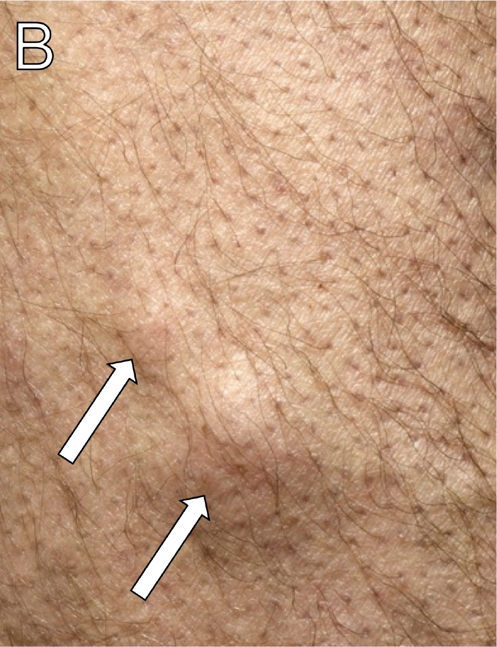
A man in his early 40s originally from Ethiopia presented with epistaxis, dizziness, weakness, and difficulty walking. He was febrile and anemic, and had several brown-black skin lesions (Figures 1A-B). Examination showed various hyperpigmented flat lesions on the chest and extremities, ranging in size from 5 mm to 30 mm, some of which were tender to palpation. An HIV test was positive, and his CD4+ count was 4 cells/µL. A biopsy of a representative cutaneous lesion showed capillary proliferation with associated neutrophilic inflammation (Figures 1C-D), and Warthin-Starry stain was positive for rod-shaped bacilli (Figures 1E-F, stained black). A serum immunoglobin G (IgG) test was positive for Bartonella hensalae and Bartonella quintana.7 A diagnosis of bacillary angiomatosis was made, and he subsequently initiated treatment with doxycycline with improvement in the cutaneous lesions.
Candidiasis
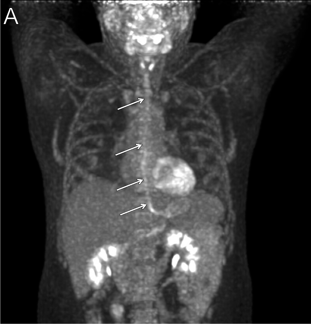
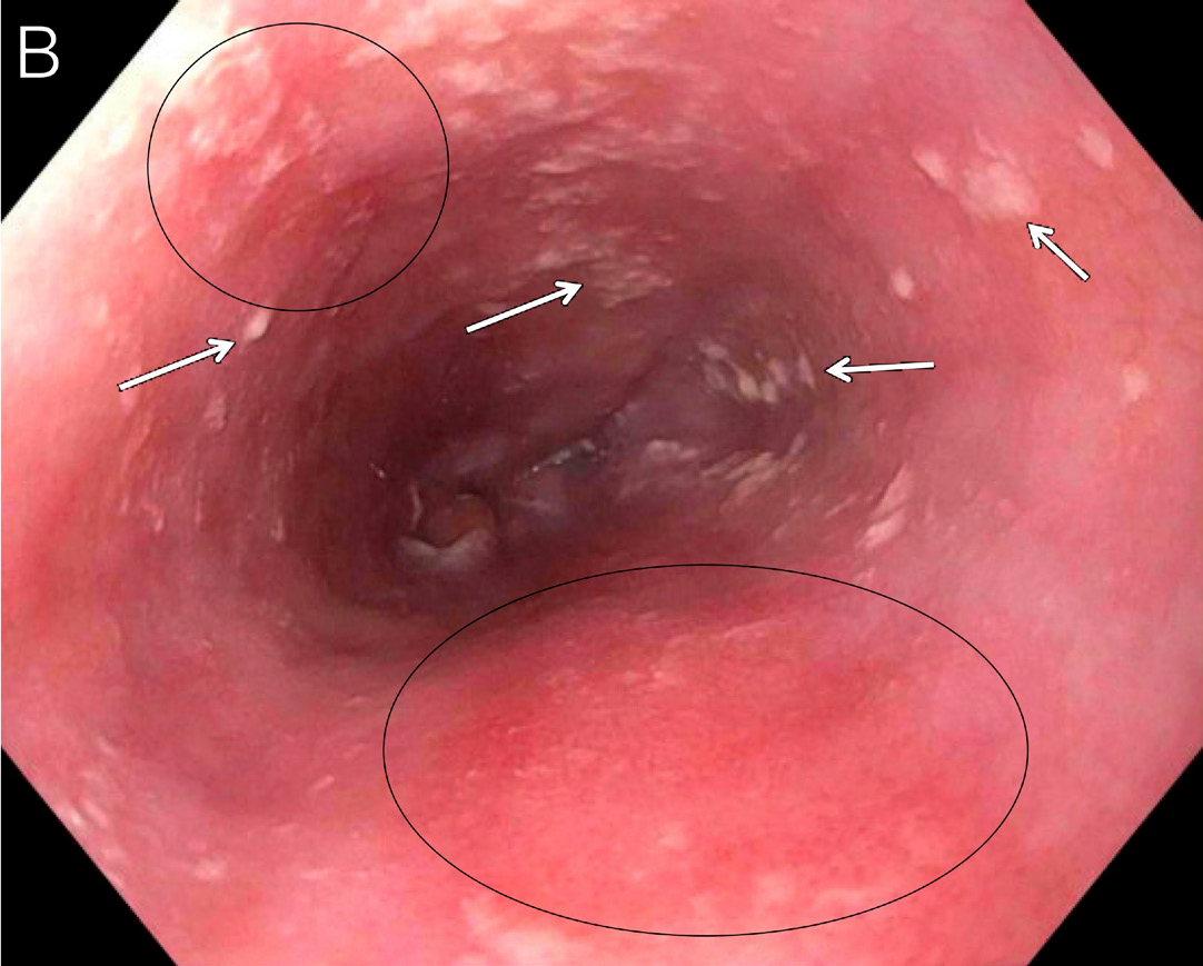
A man in his late 40s with HIV (CD4+ count, <100 cells/μL) not on ART had plasmablastic lymphoma in remission and reported dysphagia for the preceding month. Positron emission tomography (PET) computed tomography (CT) scan showed increased uptake along the entire esophagus (Figure 2A, arrows), suspicious for esophagitis. Endoscopy showed erythematous mucosa (Figure 2B, circles) and white plaques (Figure 2B, arrows) throughout the entire esophagus, concerning for esophageal candidiasis.8 He initiated intravenous (IV) micafungin followed by oral fluconazole with clinical improvement.
Cryptococcosis
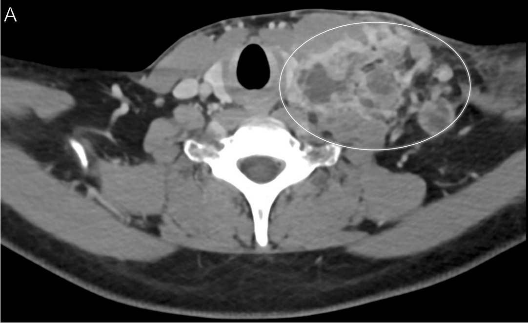
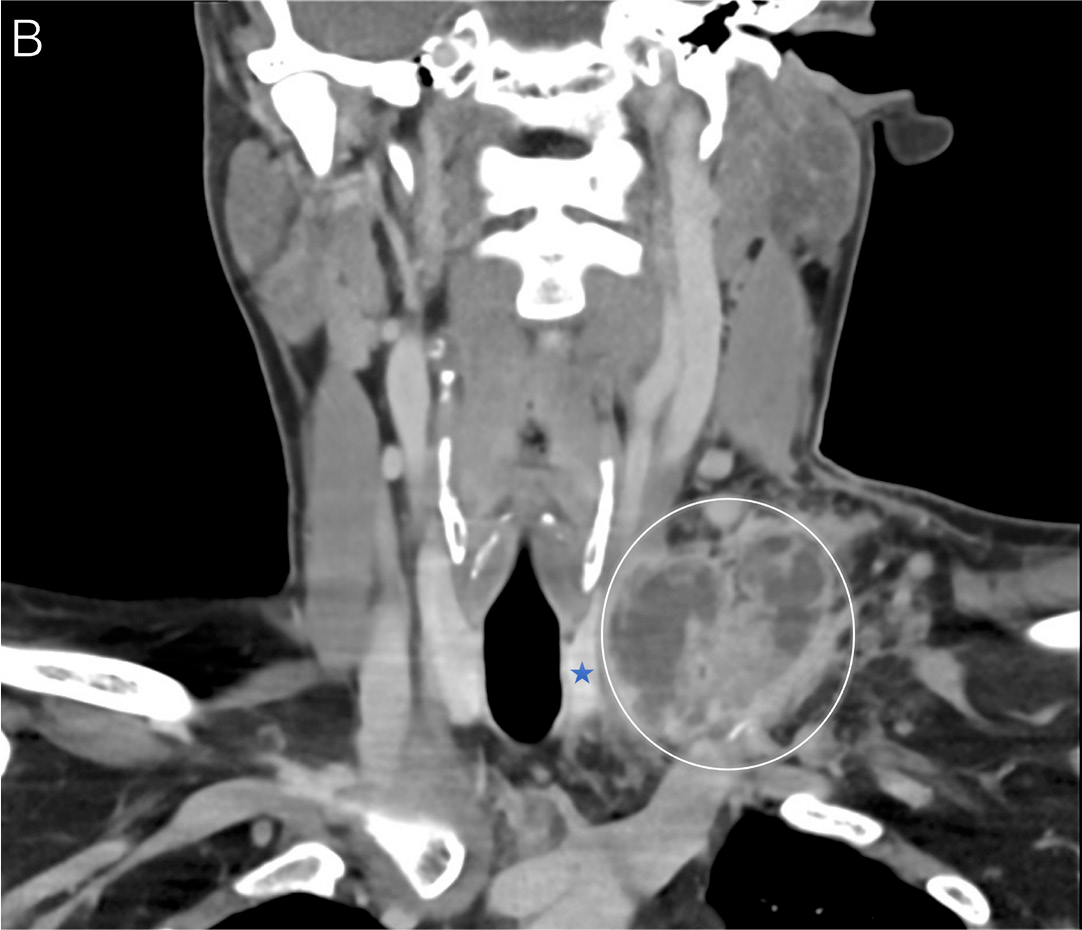
A man in his mid-30s with HIV (CD4+ count, 186 cells/μL) and cryptococcal meningitis (for which he took maintenance fluconazole) had initiated ART 1 year ago before presenting with newly developed, tender supraclavicular lymphadenopathy. A CT scan of the neck revealed an ill-defined necrotic soft tissue mass in the left supraclavicular region (Figures 3A–B, circles) measuring approximately 6 × 4 cm, displacing the thyroid gland (Figure 3B, star) slightly to the right and the left sternocleidomastoid muscle anteriorly. Biopsy of the tissue revealed epithelioid histiocytes forming granulomas, and Grocott's methenamine silver (GMS) stain was positive for oval structures consistent with Cryptococcus spp.9 He was diagnosed with cryptococcal immune reconstitution inflammatory syndrome (IRIS). He received a brief course of corticosteroids and continued maintenance fluconazole with ultimate resolution of the lymphadenopathy.
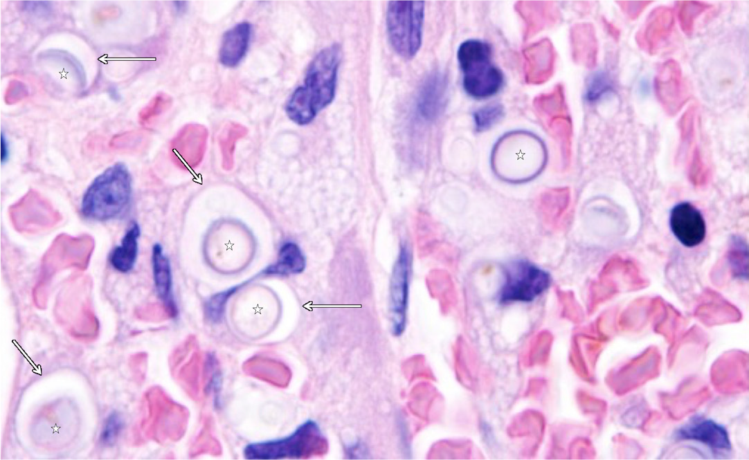
A woman in her early 60s with HIV not on ART presented with weight loss, fevers, headaches, and marked pancytopenia. Cerebrospinal fluid (CSF) cryptococcal antigen test was positive. A hematoxylin and eosin (H&E) stained section of a bone marrow biopsy revealed several poorly formed granulomas with yeast cells (Figure 4, stars) of variable size, some of which were encapsulated (Figure 4, arrows), consistent with disseminated cryptococcosis.9
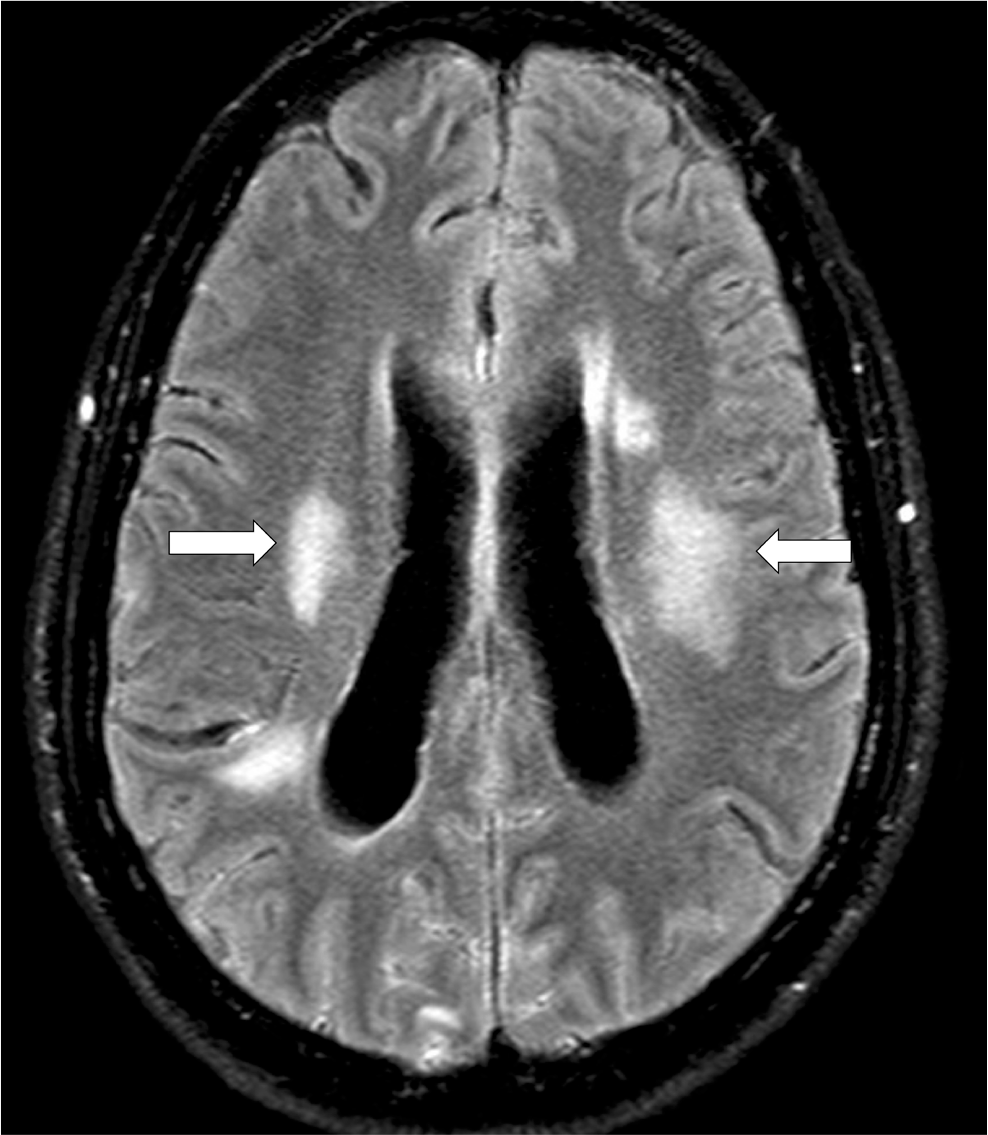
A man in his mid-40s was diagnosed with HIV (CD4+ count, 117 cells/μL) and cryptococcal meningitis. He initiated treatment for both. Seven months later, he presented with recurrence of headaches and mental status changes. Magnetic resonance imaging (MRI) of the brain showed diffuse leptomeningeal enhancement, with areas of hyperintensity in the bilateral temporal lobes (Figure 5, arrows) and parieto-occipital lobe, concerning for cryptococcal IRIS.9 He reinitiated induction therapy with amphotericin B and initiated high-dose corticosteroids. He required repeated lumbar punctures for management of increased intracranial pressure. He subsequently improved, tapered off corticosteroids, and transitioned to maintenance fluconazole.
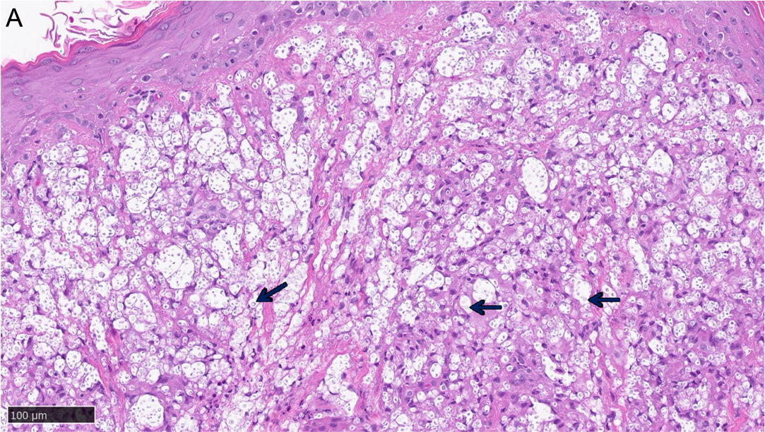
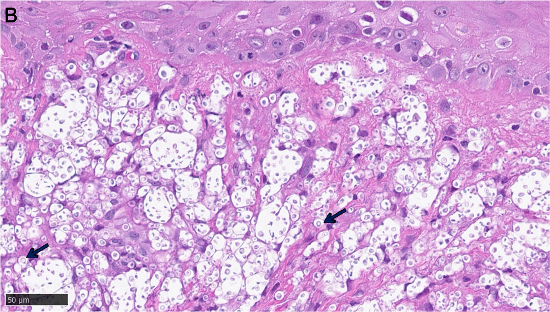
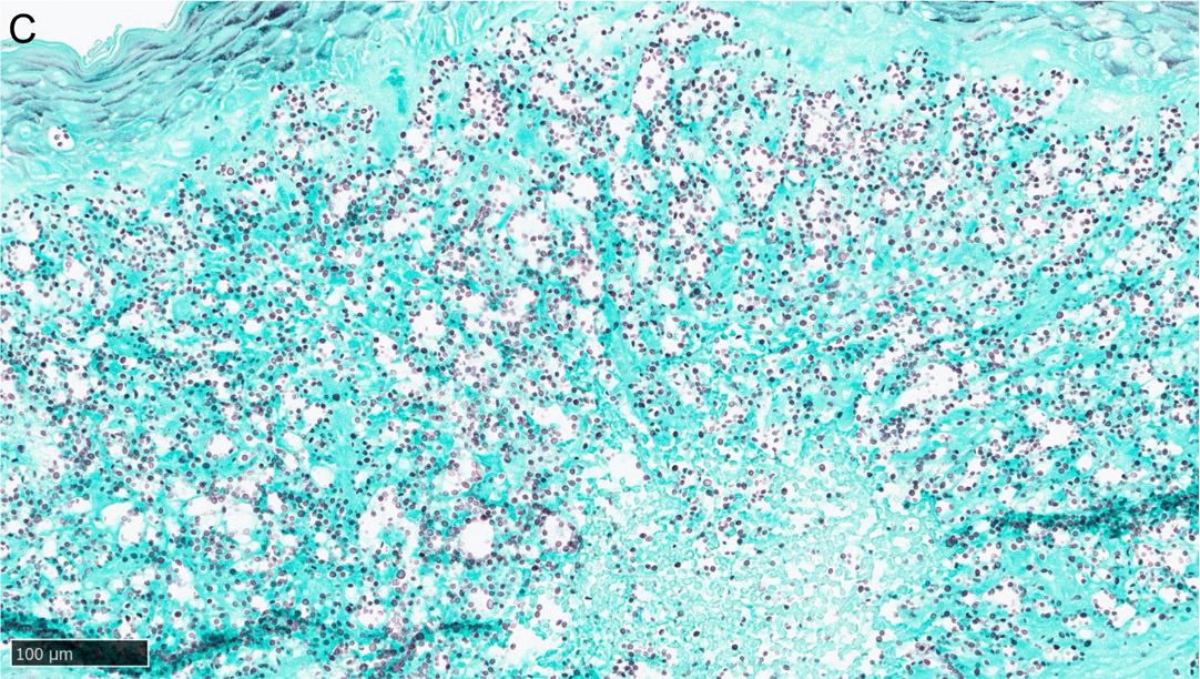
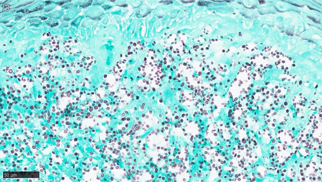
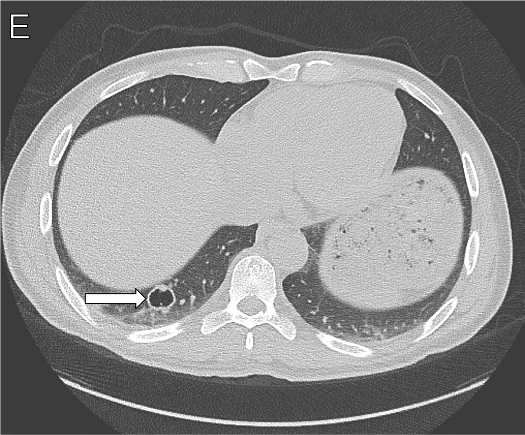
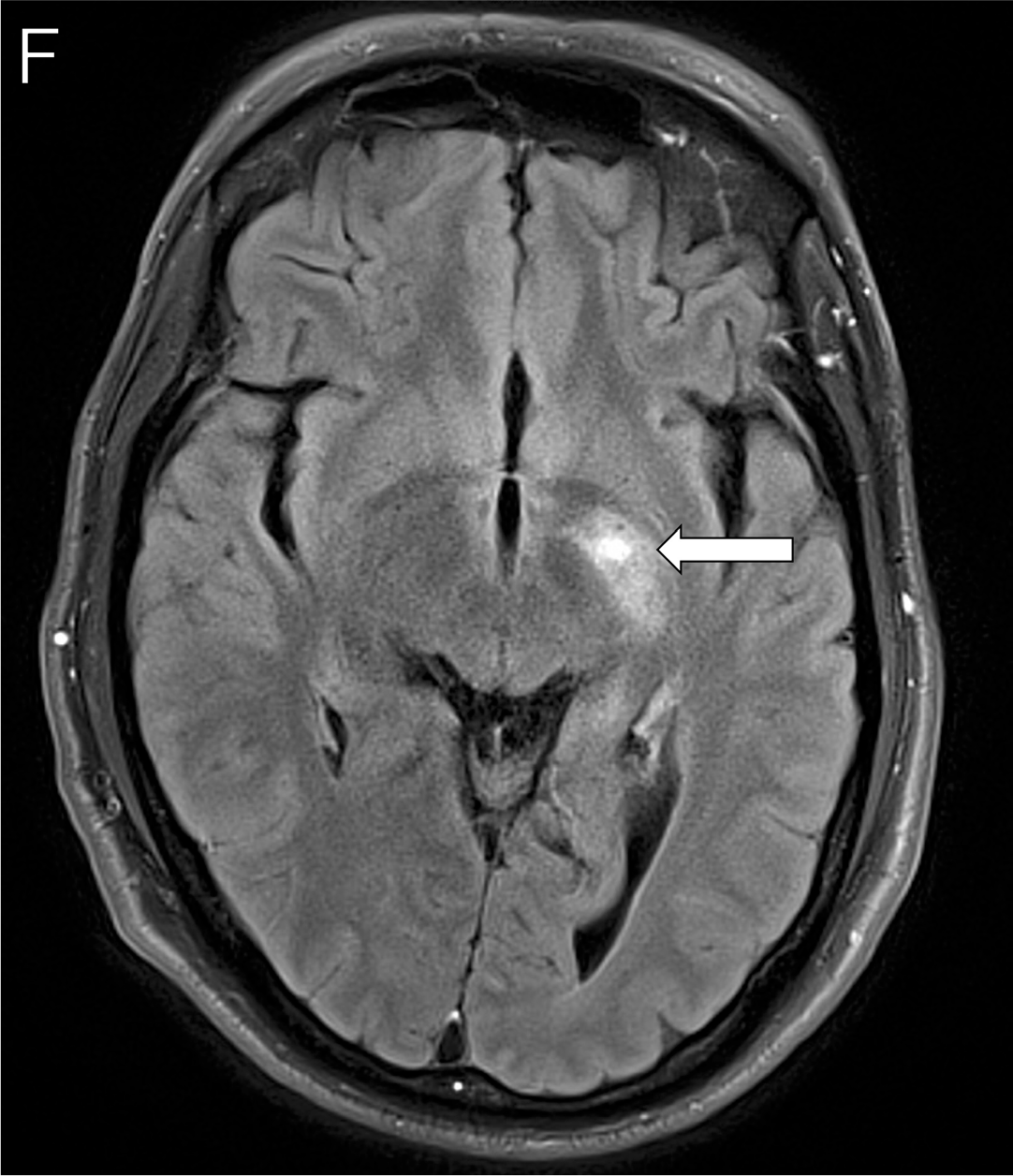
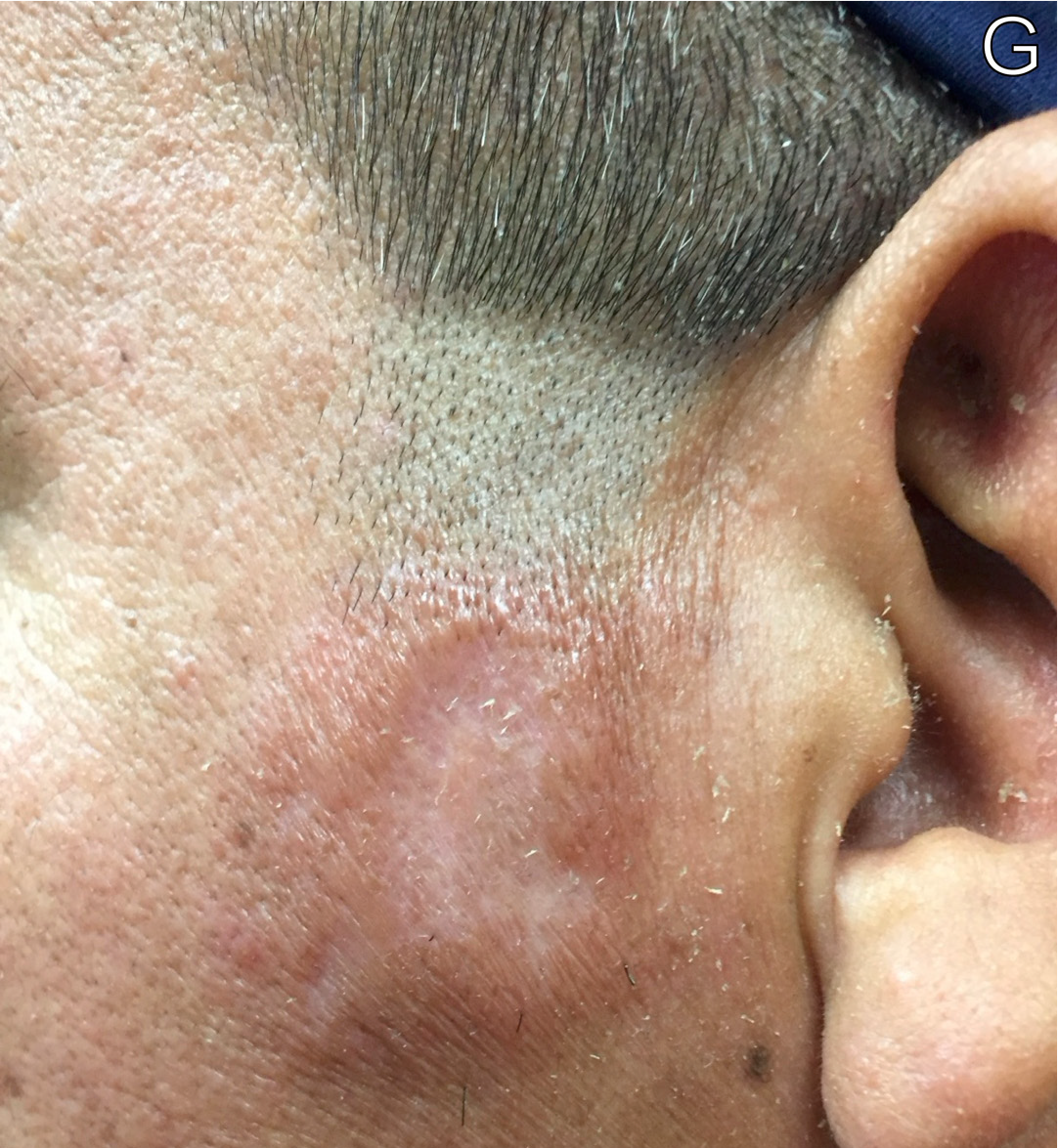
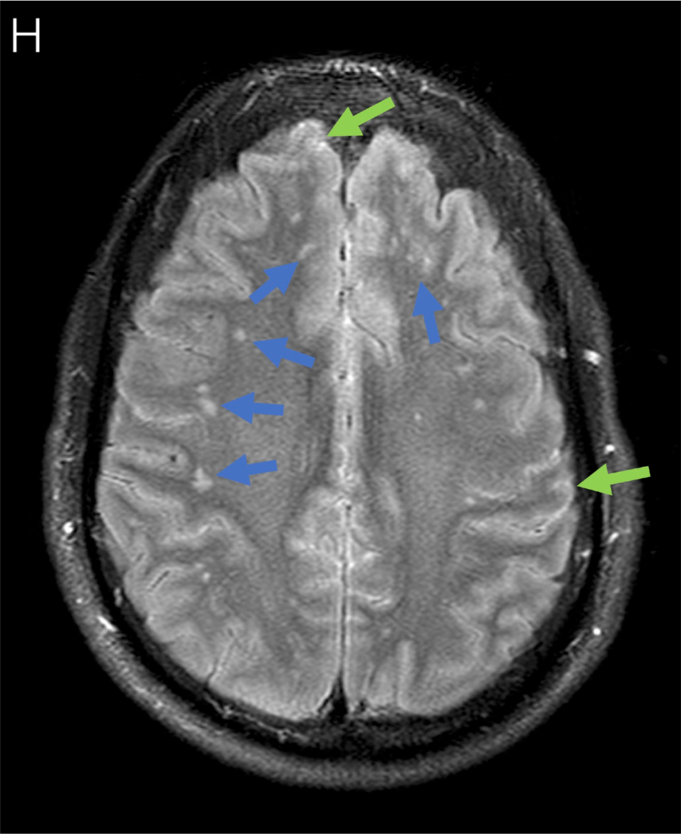
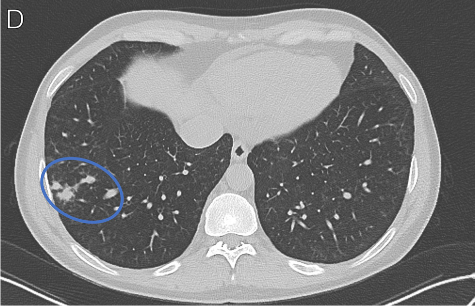
A man in his mid-40s originally from El Salvador presented to a hospital with weight loss, subjective fevers, headache, and photophobia. CSF cryptococcal antigen test was positive, and fungal cultures grew Cryptococcus neoformans. He also had a nonhealing nodular lesion with central ulceration over the left temporal region of his face. The lesion was biopsied and demonstrated infiltration of the entire dermis with round fungal elements, as shown in H&E (Figures 6A–B, arrows) and GMS stains (Figures 6C-D, stained black), consistent with Cryptococcus spp infection. An HIV test was positive, and his CD4+ count was 10 cells/μL. He initiated treatment with amphotericin B and flucytosine followed by fluconazole. A CT scan of the chest demonstrated cavitary nodules within the right lung base (Figure 6E, arrow). After 2 weeks of fluconazole and negative repeat CSF cultures, he initiated ART. About 3 months later, he redeveloped head and neck pain, and an MRI of the brain revealed a new enhancing lesion in the left basal ganglia (Figure 6F, arrow). CSF studies did not reveal a new infectious process, consistent with IRIS secondary to cryptococcal disease. He initiated corticosteroids with improvement in his symptoms, and then tapered off corticosteroids over a 2-month period. About 3 months after stopping corticosteroids, he redeveloped headaches and subjective fevers, and the left temporal skin lesion had become more prominent (Figure 6G). He reinitiated amphotericin B while further work-up was pending. Repeat MRI of the brain showed various new periventricular and subcortical white matter hyperintensities (Figure 6H, blue arrows) and interval increase in leptomeningeal enhancement (Figure 6H, green arrows). Repeat skin biopsy showed inflammatory infiltrate and cryptococcal elements, and CSF studies demonstrated ongoing inflammation. Cultures from the sites were negative, consistent with recurrent cryptococcal IRIS. Amphotericin B was discontinued, and he reinitiated fluconazole and corticosteroids. His symptoms improved, the skin lesion flattened, and corticosteroids were gradually tapered.
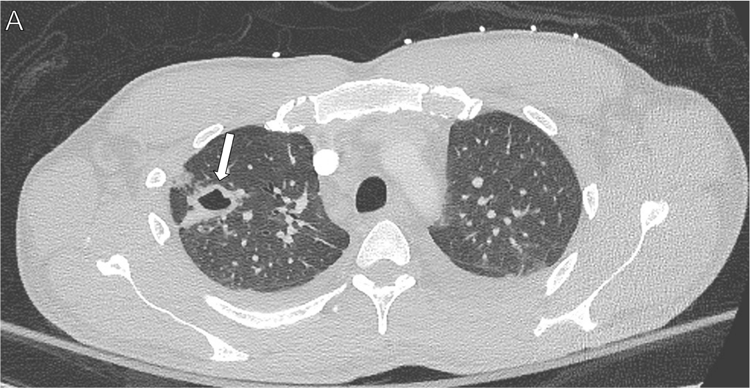
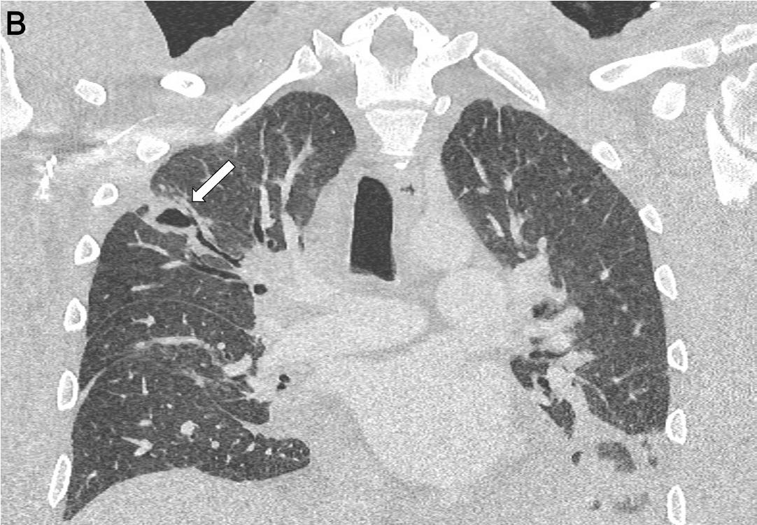
A man in his mid-30s was diagnosed with HIV (CD4+ count, 106 cells/μL) and initiated ART. He presented to the hospital 2 weeks later with a 3-day history of fever and fatigue. A CT scan of the chest revealed a cavitary pulmonary lesion (Figures 7A-B, arrows) and paratracheal lymphadenopathy. He was started empirically on tuberculosis (TB) therapy as well as therapy for Pneumocystis pneumonia and bacterial pneumonia, but he remained persistently febrile and tachycardic and became hypotensive. Bronchoscopy demonstrated normal bronchial mucosa with no endobronchial lesions, and copious thin, watery secretions. Bronchoscopy fungal culture grew Cryptococcus neoformans. He was treated with liposomal amphotericin for 1 week and then transitioned to oral fluconazole with improvement in his clinical status.9,10
Cytomegalovirus
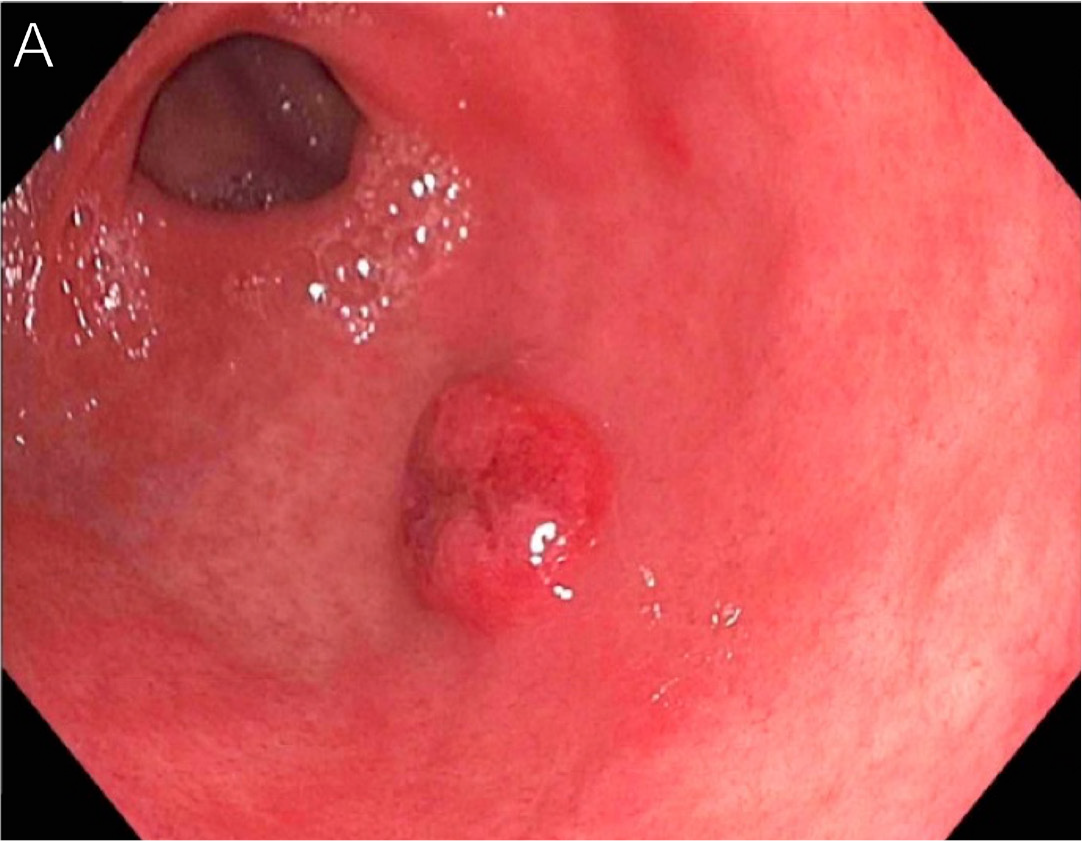
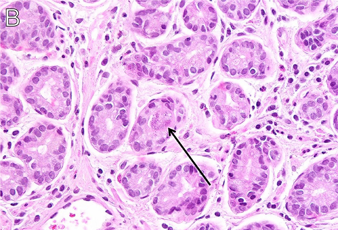
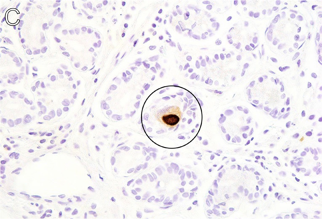
A man in his early 20s with HIV (CD4+ count, 10 cells/μL) not on ART presented with postprandial vomiting, abdominal pain, and diarrhea. Upper endoscopy revealed a medium-sized (8 mm), sessile, noncircumferential mass in the gastric body and mildly erythematous mucosa (Figure 8A). Anatomic pathology showed cytopathic effect in hematoxylin and eotaxin stain (Figure 8B, arrow) and positive cytomegalovirus (CMV) immunostain (Figure 8C, circle). He was diagnosed with CMV gastritis and initiated valganciclovir with improvement in his symptoms.11
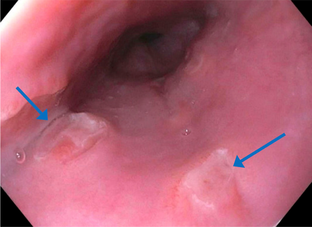
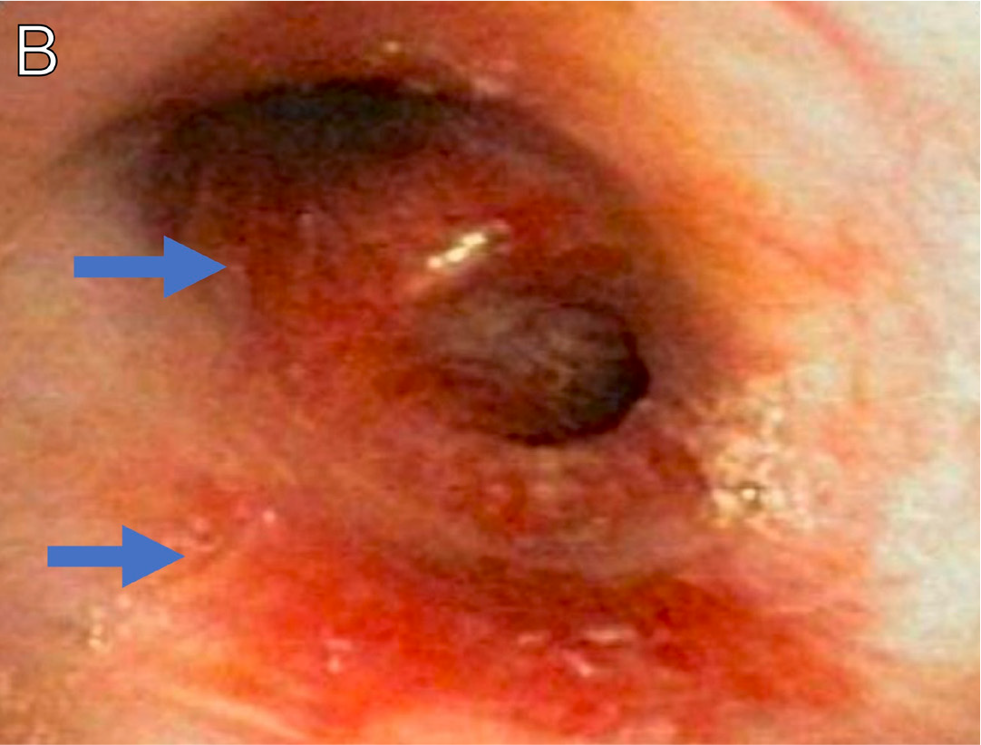
A man in his early 60s with HIV (CD4+ count, 27 cells/μL) not on ART presented with weight loss and substantial odynophagia refractory to fluconazole treatment. Upper endoscopy revealed a few cratered esophageal ulcers with stigmata of recent bleeding in the midesophagus, with the largest lesion measuring 5 mm (Figure 9, arrows). Pathology showed intramucosal neutrophilic inflammation and positive CMV immunohistochemistry stain. He was diagnosed with CMV esophagitis and initiated valganciclovir with improvement in his symptoms.11
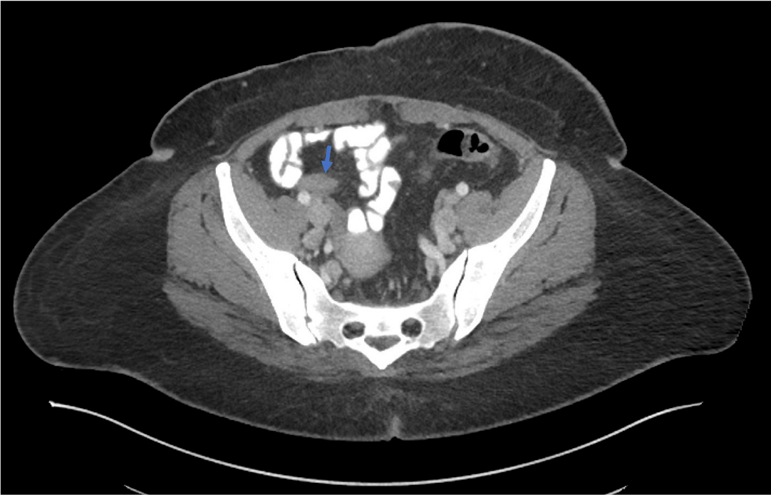
A woman in her 30s initially presented with pharyngitis and dysphagia. She was diagnosed with HIV (CD4+ count, 72 cells/µL) and oral candidiasis, and initiated ART. Two weeks after ART initiation, she presented with right lower abdominal pain. CT imaging revealed appendiceal enlargement and mild stranding consistent with early appendicitis (Figure 10, arrow). She underwent laparoscopic appendectomy, and pathology revealed findings consistent with acute appendicitis and positive CMV immunostain. She had subsequent improvement following surgery; however, approximately 3 weeks later, she presented with hematochezia. A decision to pursue colonoscopy was deferred due to recent appendectomy. She was treated empirically for CMV colitis with valganciclovir, with subsequent improvement.11,12
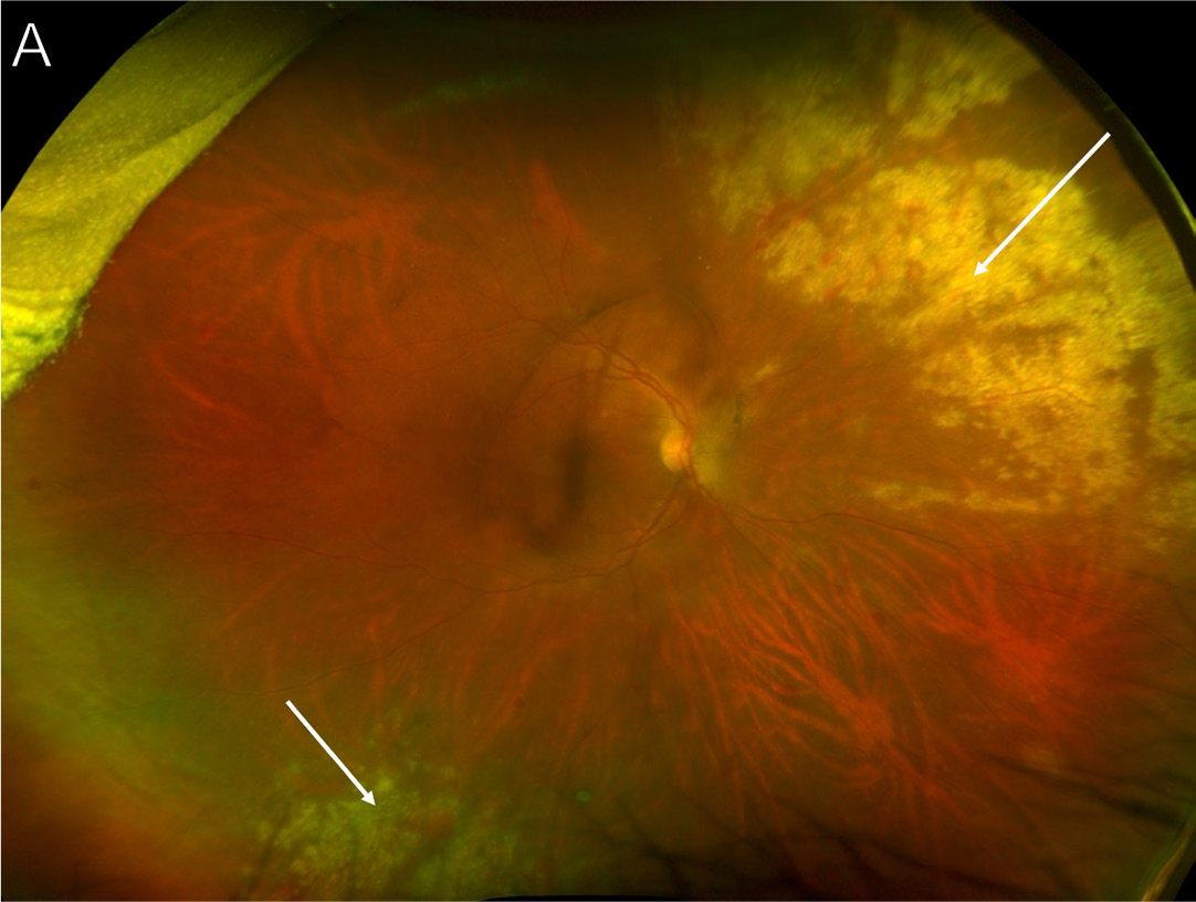
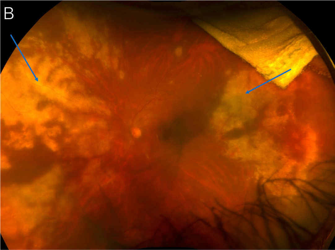
A man in his early 50s with HIV (CD4+ count, 4 cells/μ L) not on ART presented with left eye floaters for the preceding 3 weeks. A blood CMV polymerase chain reaction (PCR) test was positive. Dilated eye exam revealed right eye peripheral retinitis (Figure 11A, arrows) with patchy hemorrhage, as well as left eye vitritis and extensive peripheral retinitis (Figure 11B, arrows) with scattered hemorrhage encroaching toward the macula and optic nerve, consistent with bilateral CMV retinitis.11
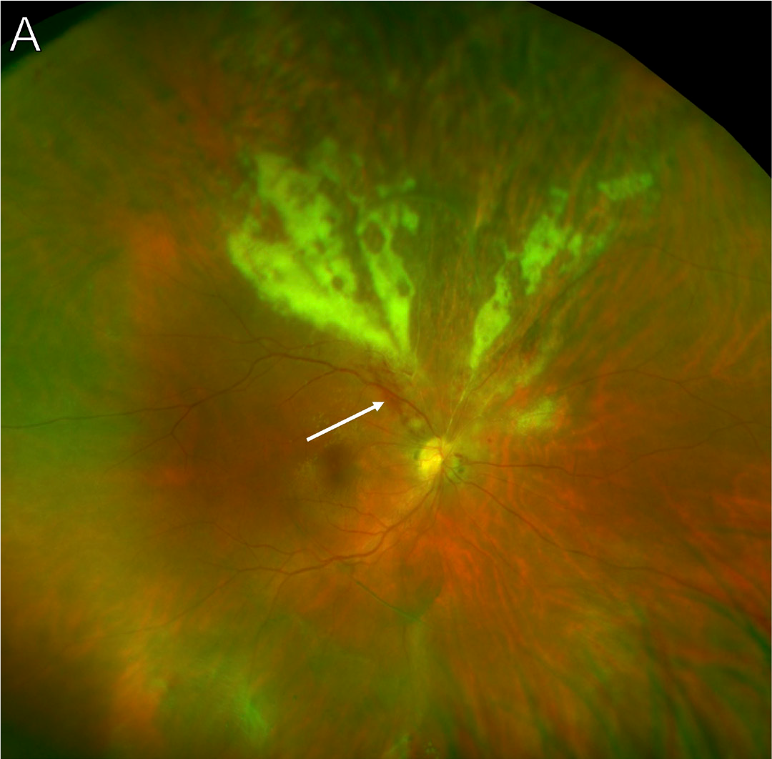
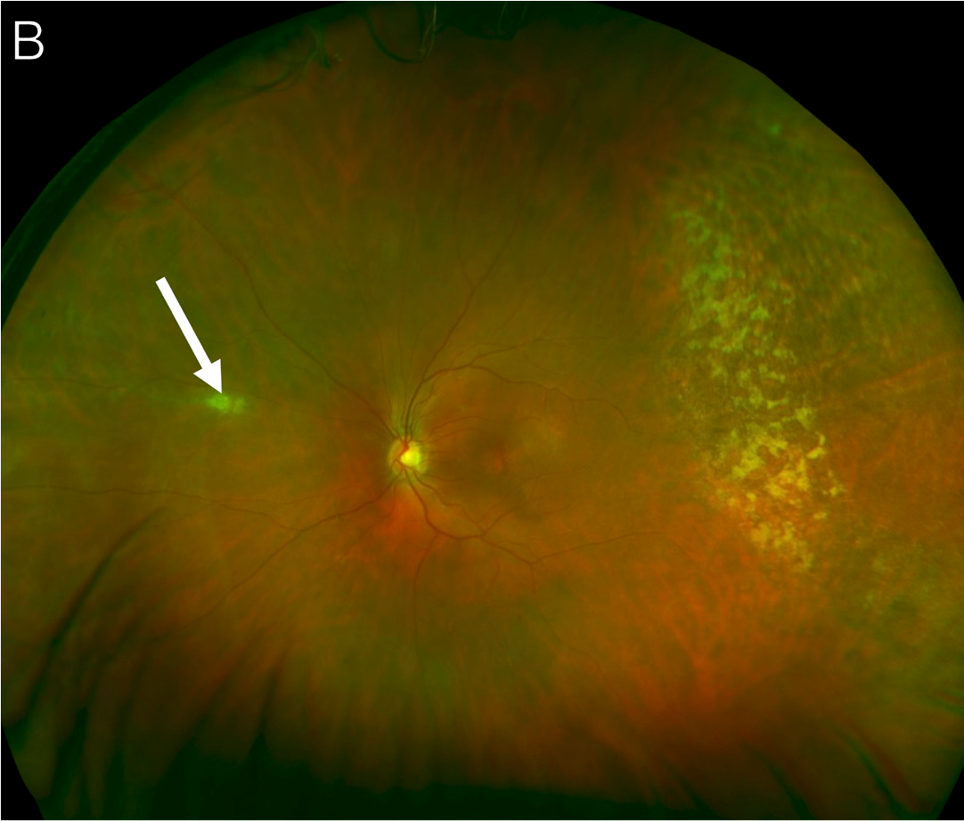
A man in his late 30s with advanced HIV not on ART presented with a 1- to 2-year history of right eye floaters. Dilated eye exam showed right eye wedge-shaped retinitis superior to the optic disc with associated hemorrhage (Figure 12A, arrow), and left eye granular-type retinitis in the temporal periphery (3:00) as well as a lesion nasally (Figure 12B, arrow), consistent with bilateral CMV retinitis.11
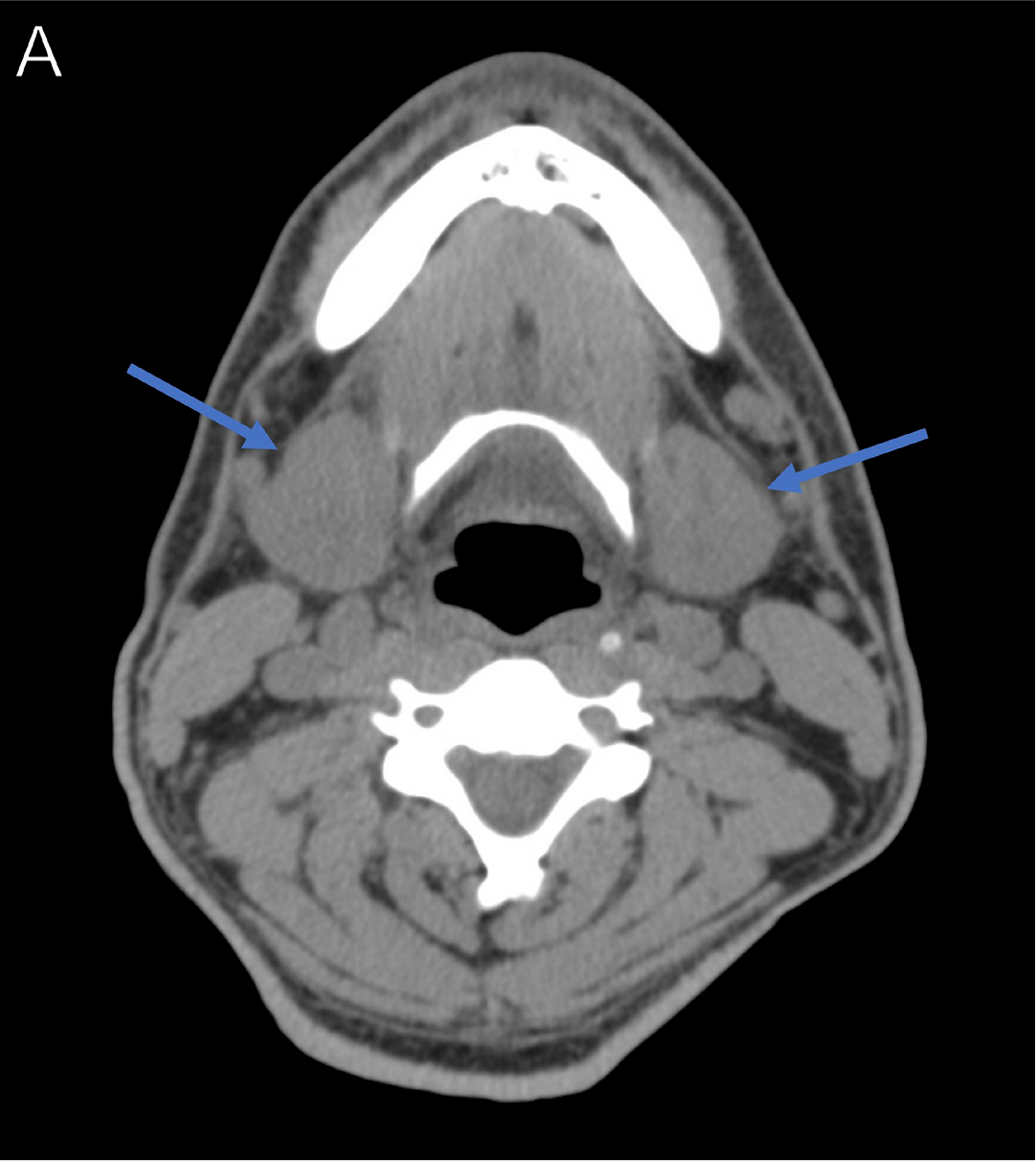
A man in his early 40s with HIV (CD4+ count, 4 cells/μL) developed fever and acute onset left neck swelling and pain 4 weeks after ART initiation. Physical exam revealed mild, hard, nontender swelling in the submandibular region predominantly on the left. A CT scan showed bilateral enlargement of the major salivary glands (Figure 13A, arrows). Symptoms were refractory to treatment with amoxicillin/clavulanate. Fine-needle aspiration was performed and showed positive CMV immunostain with cytopathic changes (Figures 13B-C), consistent with CMV sialadenitis.11,13
Epstein-Barr Virus
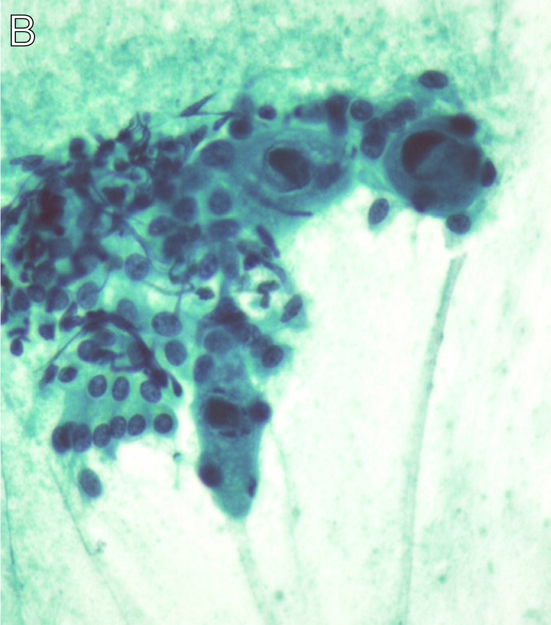
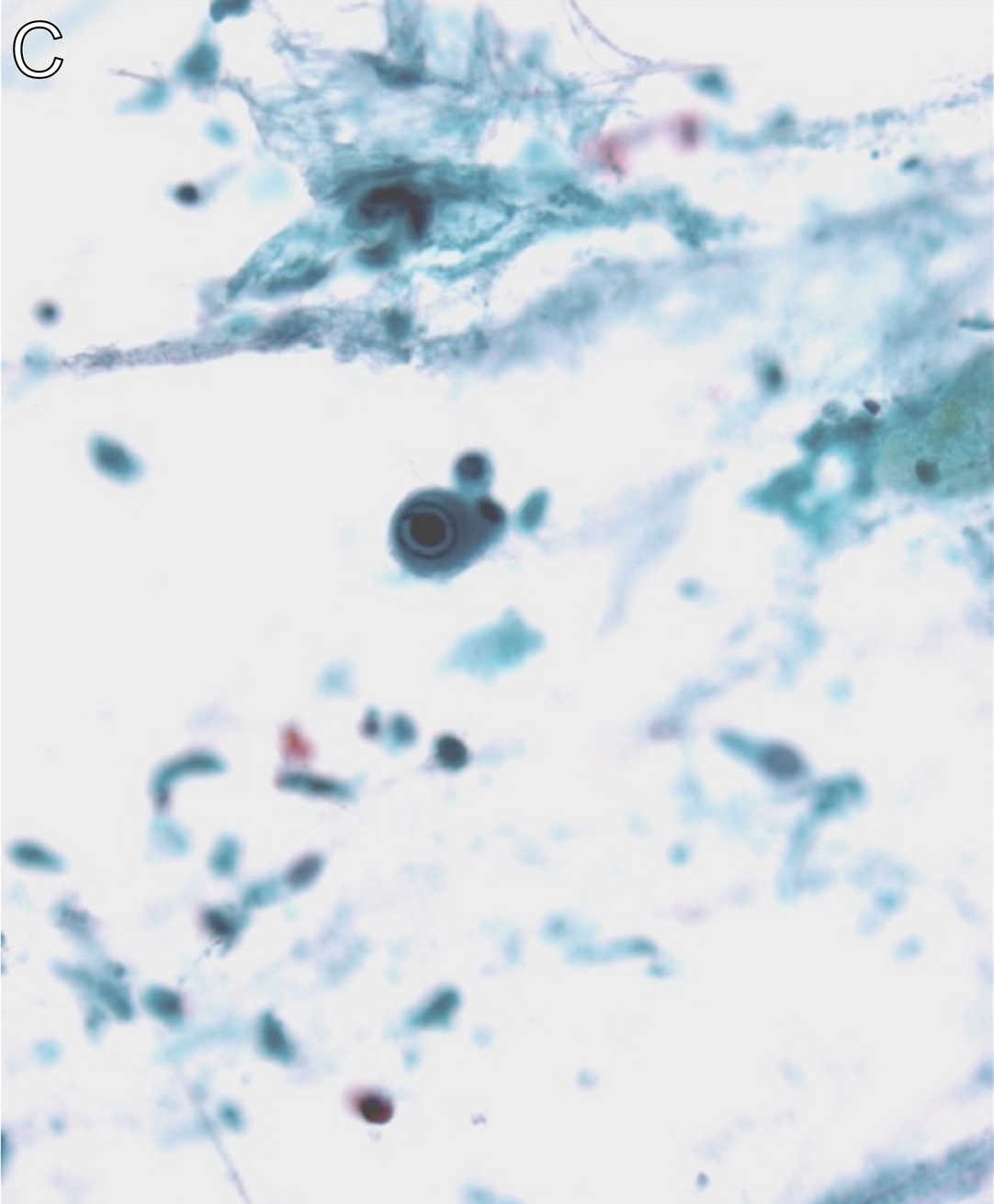
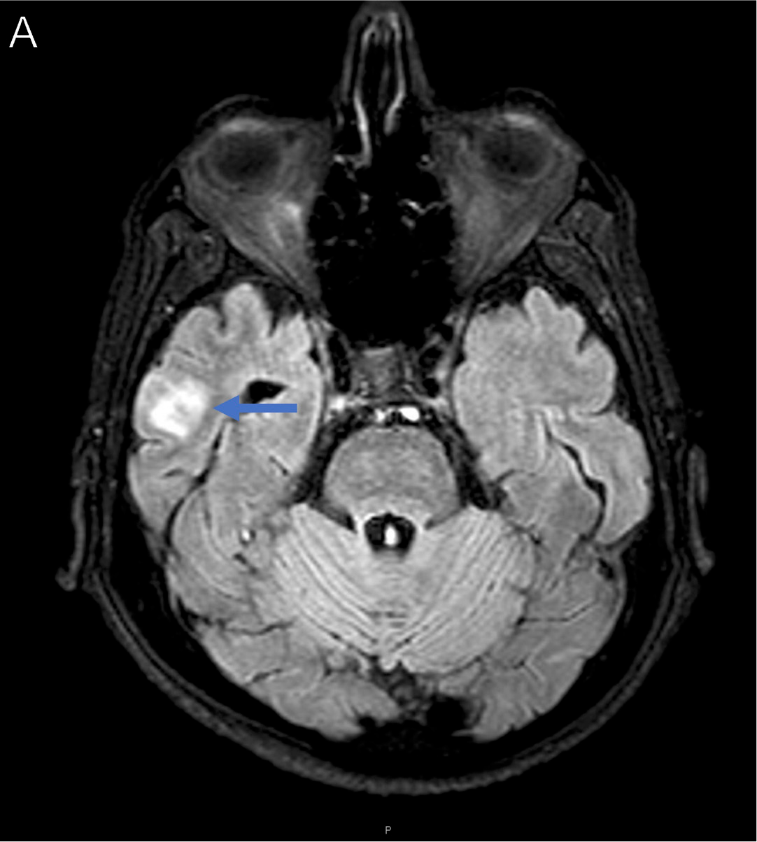
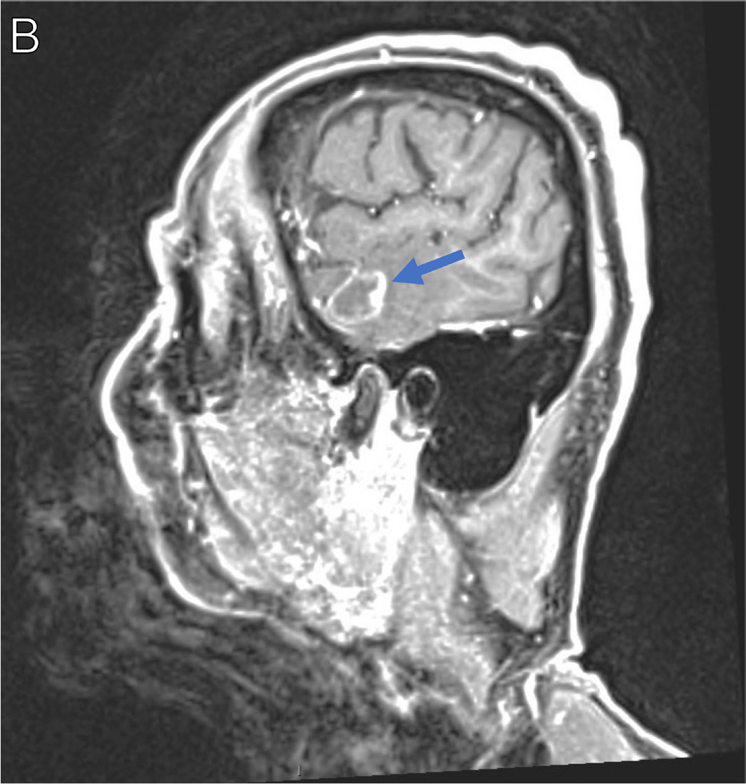
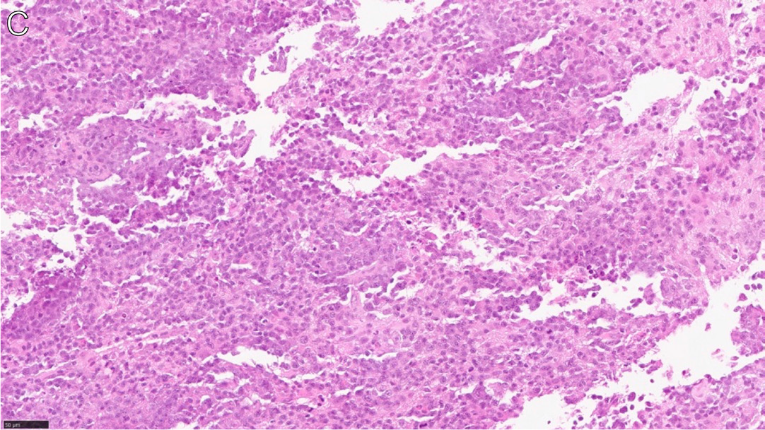
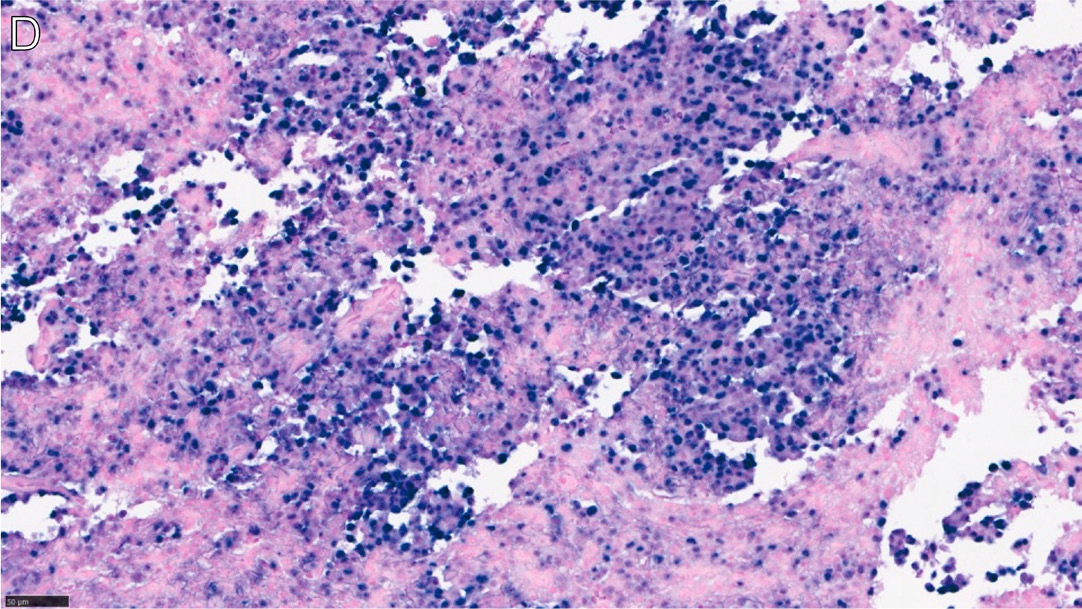
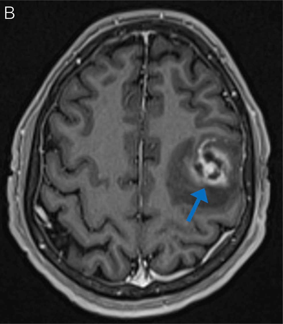
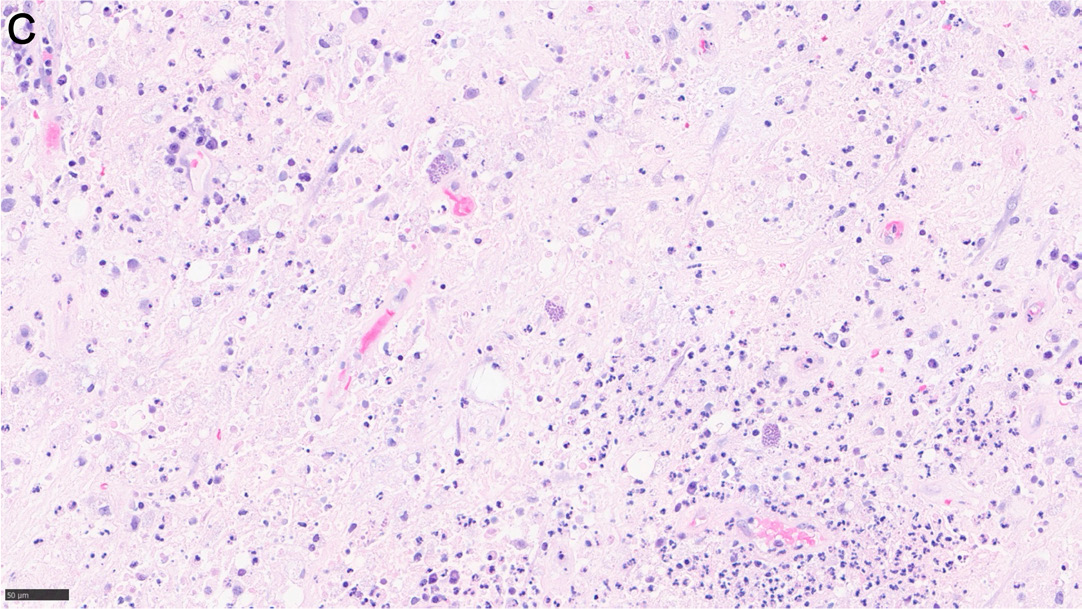
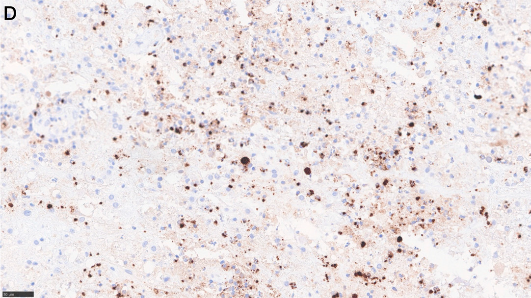
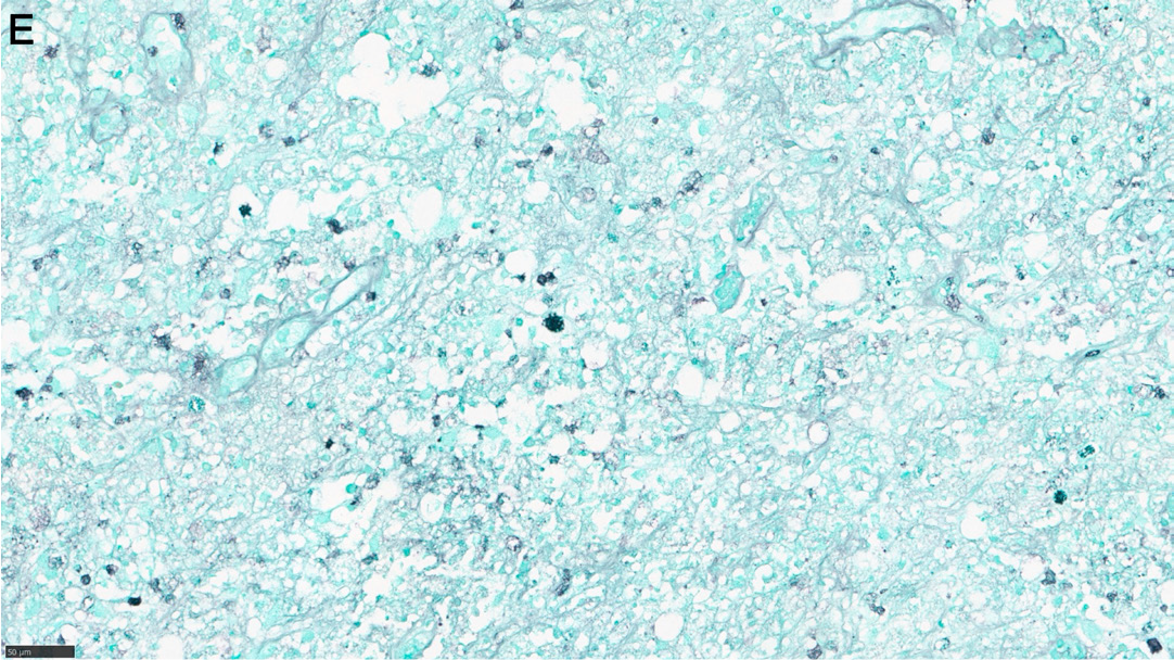
A man in his early 60s with advanced HIV (CD4+ count, 27 cells/μL) recently started ART and was incidentally found to have a focus of increased T2-weighted fluid-attenuated inversion recovery (T2/FLAIR) signal on a noncontrast brain MRI (Figure 14A, arrow). He started on treatment for toxoplasmosis; however, repeat brain MRIs with contrast over the next 6 weeks showed gradual enlargement in the size of the lesion (Figure 14B, arrow). Brain biopsy was pursued, and showed large, atypical lymphoid cells of B-cell lineage with extensive necrosis (Figure 14C), and positive Epstein-Barr virus (EBV)-encoded RNA (EBER) in situ hybridization (Figure 14D), consistent with EBV-positive diffuse large B-cell lymphoma. He began treatment with high-dose methotrexate and rituximab and achieved complete remission.14
Histoplasmosis
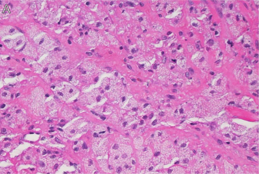
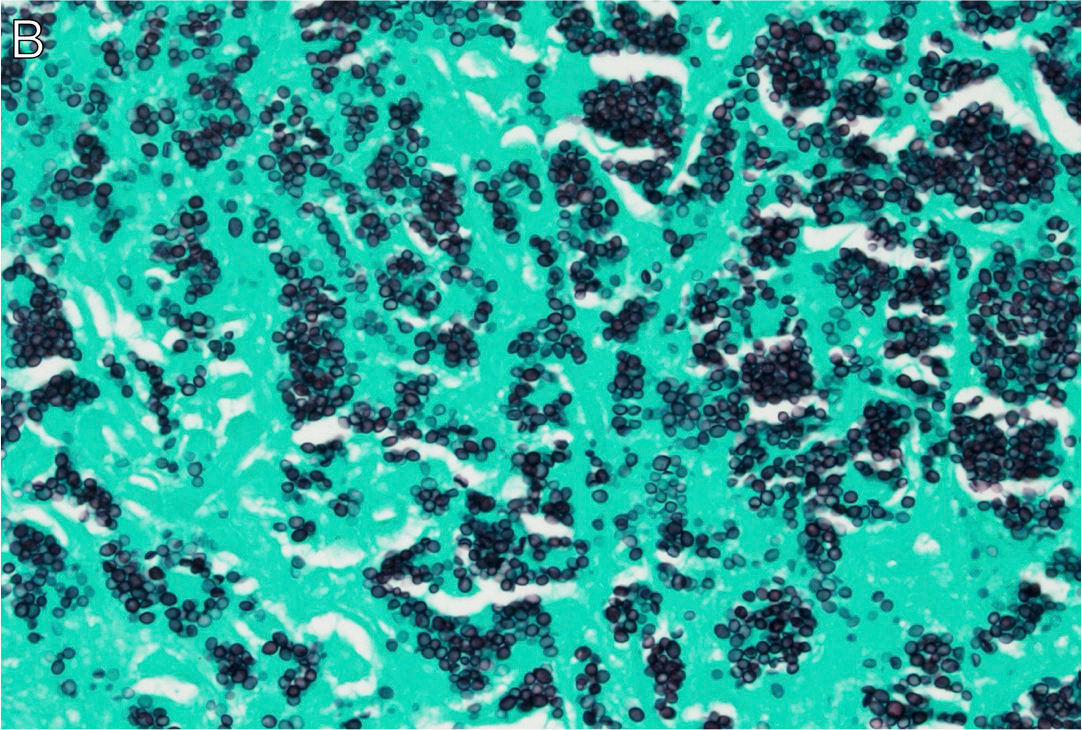
A woman in her mid-40s presented with fatigue and light-headedness. She was diagnosed with HIV (CD4+ count, 0 cells/µL) and profound pancytopenia. Bone marrow biopsy showed macrophages with atypical vacuolar cytoplasmic inclusions with positive periodic acid Schiff (PAS) and GMS stains, as well as positive urine and blood Histoplasma antigen tests.
Cervical neck lymph node biopsy showed histiocytic proliferation (Figure 15A) with massive intracellular yeast accumulation, morphologically consistent with histoplasmosis (Figure 15B). She began treatment with amphotericin B followed by itraconazole, and initiated ART.15 Her treatment course was complicated by IRIS, for which she required repeated courses of corticosteroids.
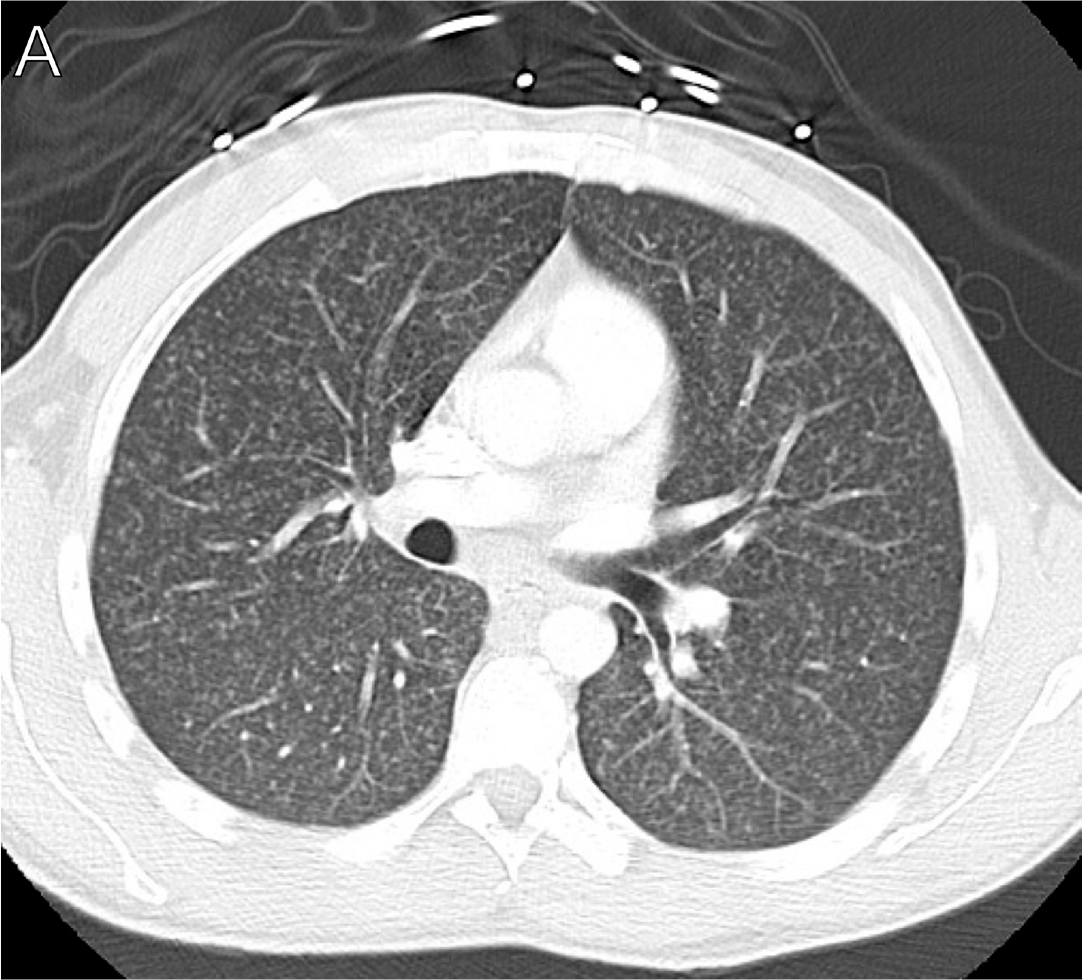
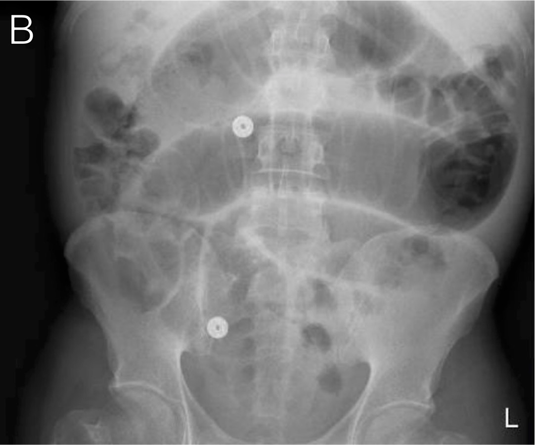
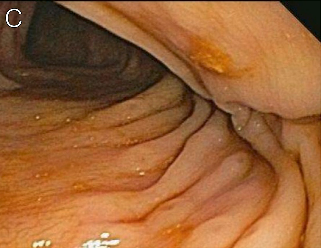
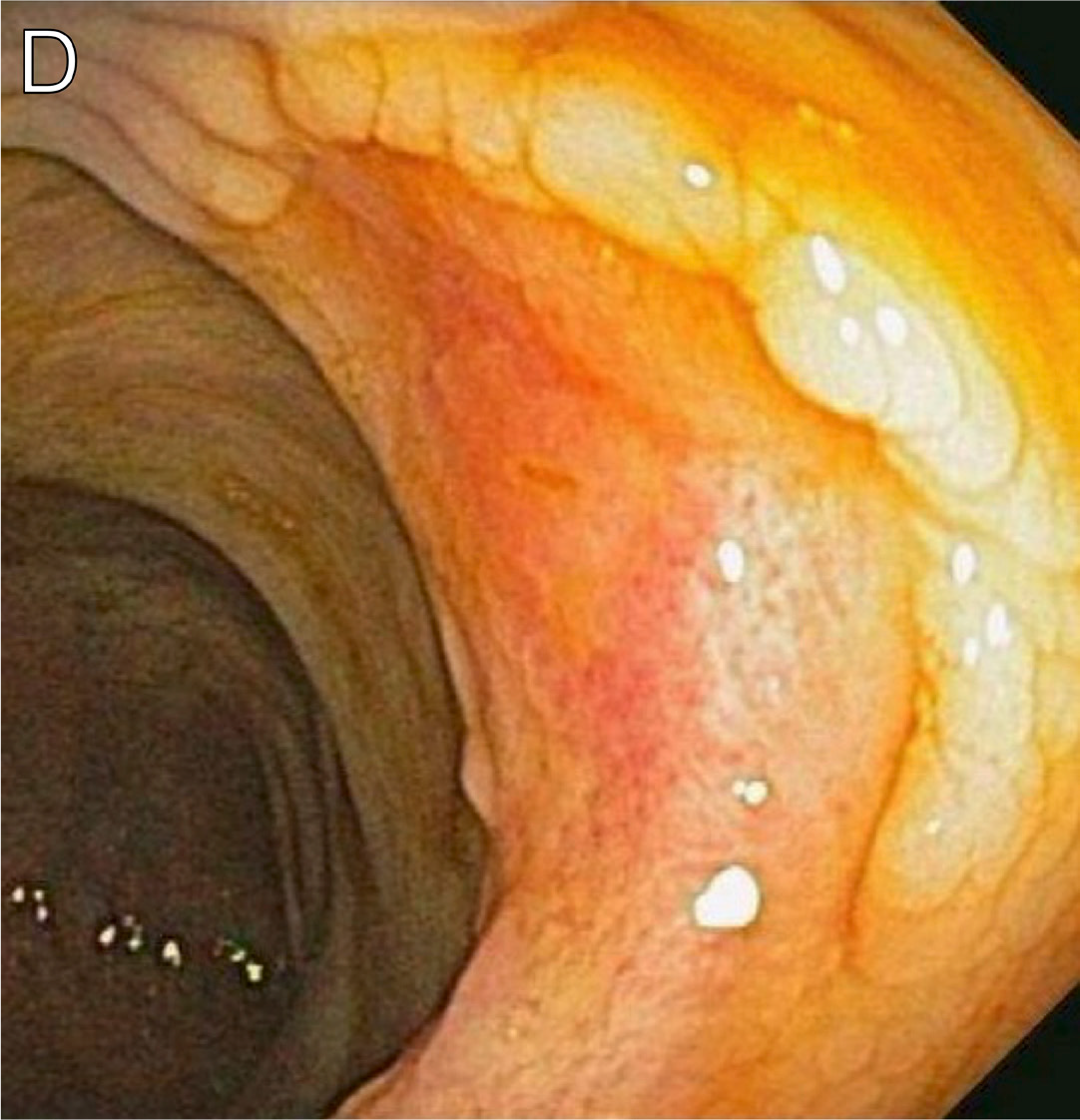
A man in his late 30s originally from Guatemala presented with abdominal pain and hematochezia. He was diagnosed with HIV and had CD4+ count of 27 cells/µL. Chest imaging demonstrated miliary stippling (Figure 16A), and abdominal imaging demonstrated an inflammatory process at the right lower quadrant involving cecum, appendix, and terminal ileum (Figure 16B). Colonoscopy revealed small, crater-like lesions in the ascending colon (Figure 16C) as well as an area of erythematous, thickened mucosa consistent with partially healed ulcer (Figure 16D). Pathology revealed Histoplasma spp, and urine Histoplasma antigen test was positive. He was treated with amphotericin B followed by itraconazole, and initiated ART. His treatment course was further complicated by partial small bowel obstruction and perforation in the setting of Histoplasma IRIS.15,16
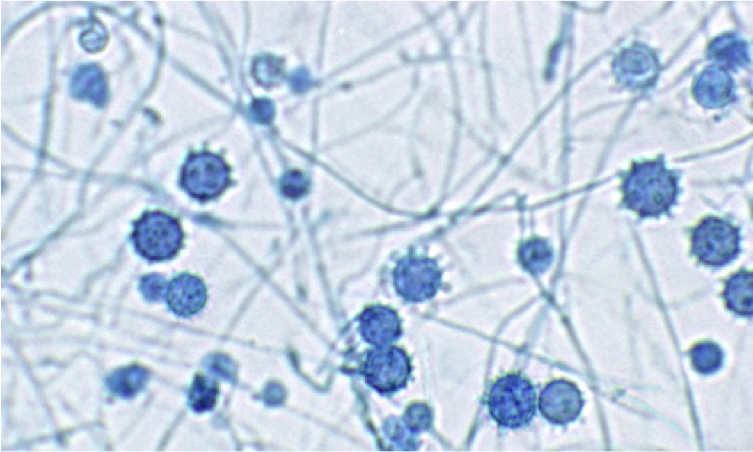
A man in his late 20s presented to the hospital with progressive lower- and upper-extremity weakness, headache, weight loss, and cough. A CT scan of the brain revealed hemorrhagic strokes in various vascular distributions. An HIV test was performed and was positive. Cultures from the bone marrow, blood, and an oral ulcer all ultimately grew Histoplasma capsulatum. Staining with lactophenol cotton blue revealed septate, hyaline hyphae with tuberculate macroconidia (Figure 17).15,16 He initiated treatment for disseminated histoplasmosis with amphotericin B followed by itraconazole. A lumbar puncture revealed positive CSF Toxoplasma gondii PCR. He was treated for CNS toxoplasmosis with pyrimethamine and sulfadiazine with improvement in his neurologic symptoms, although CNS involvement of histoplasmosis could not be definitively excluded.
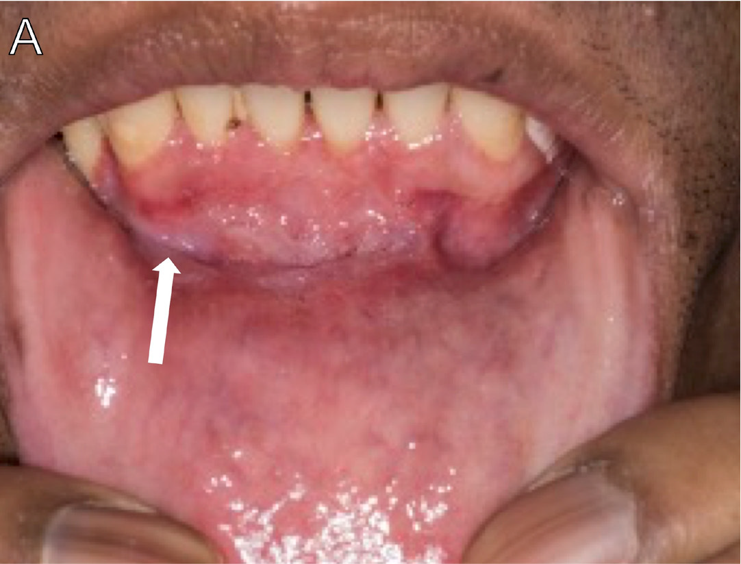
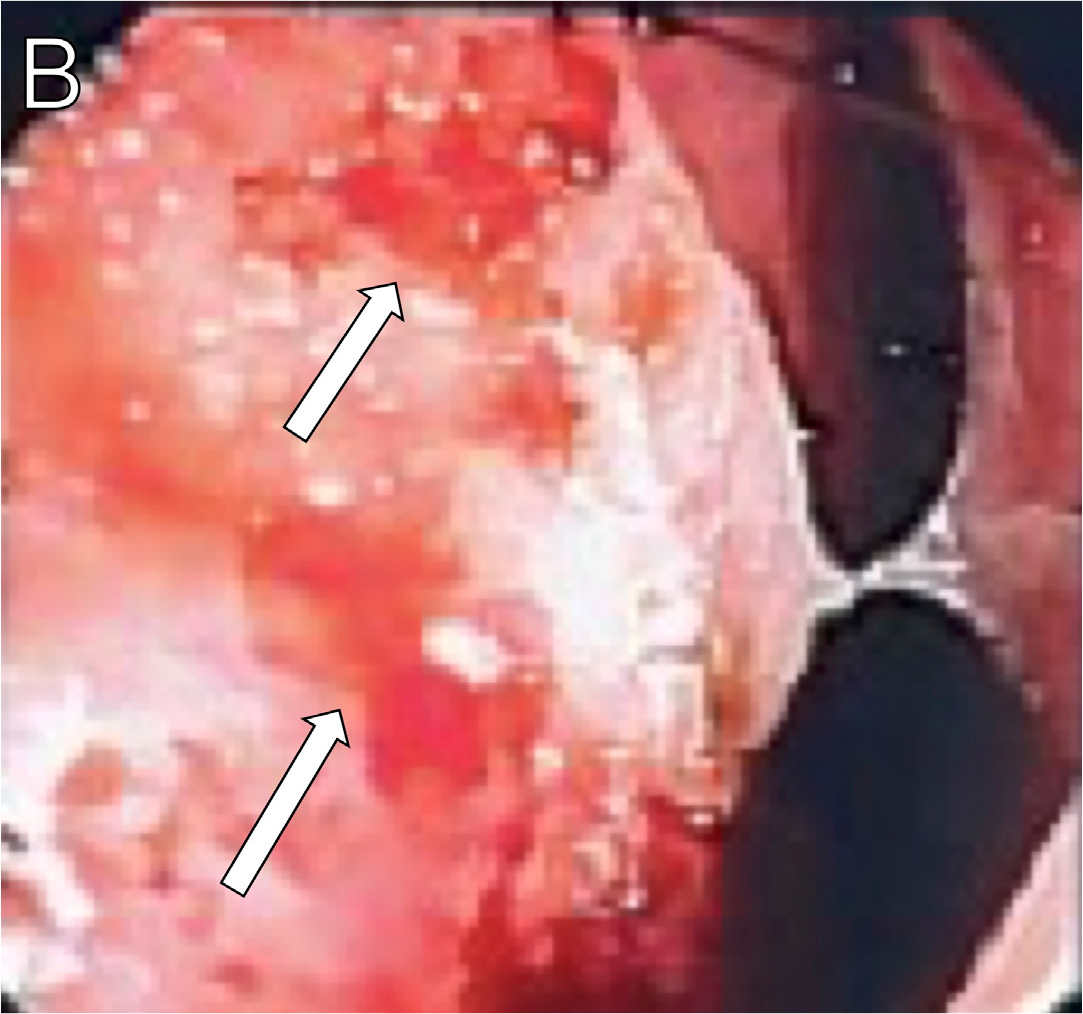
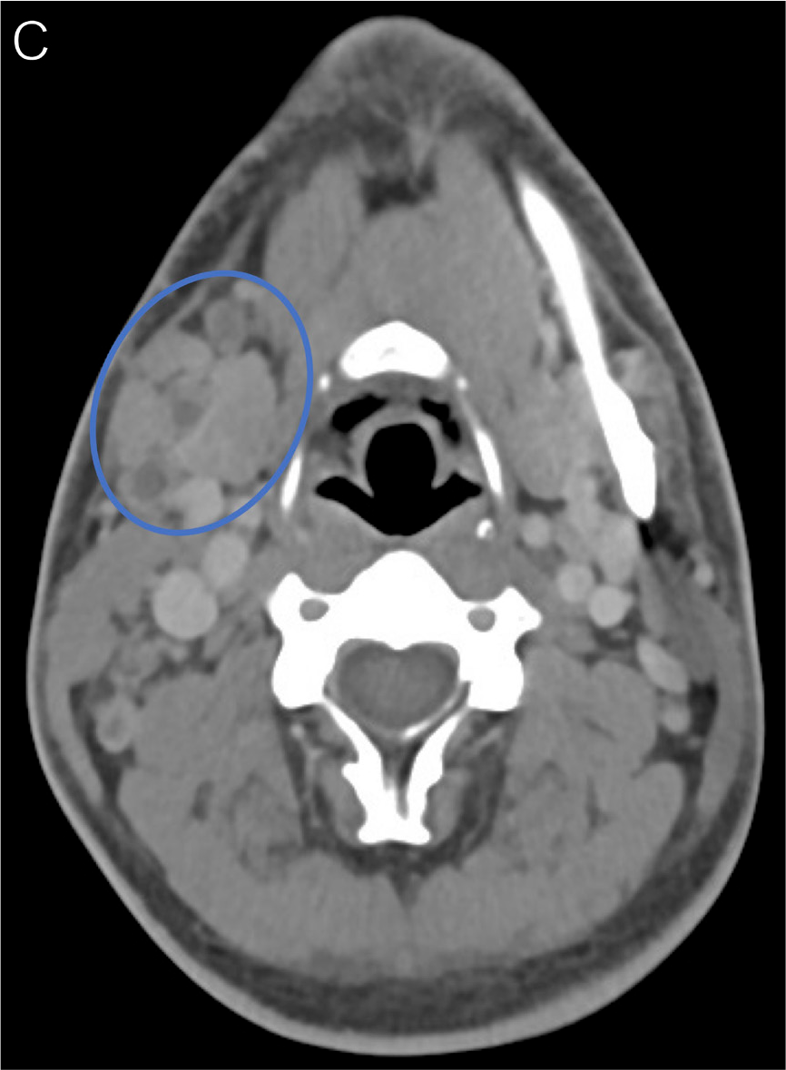
A man in his mid-30s presented with a 4-month history of fatigue, oral (Figure 18A, arrow) and nasal (Figure 18B, arrows) mucosal ulcerations, and nonhealing nodular lesions on his left elbow. He was subsequently diagnosed with HIV. An oral ulcer was biopsied and demonstrated organisms consistent with histoplasmosis. CT imaging showed extensive right-sided cervical lymphadenopathy with inflammatory stranding in the adjacent investing fatty tissues (Figure 18C, circle), and PET-CT imaging showed hypermetabolic lymph nodes above and below the diaphragm (Figure 18D, circle),15,16 consistent with disseminated histoplasmosis.
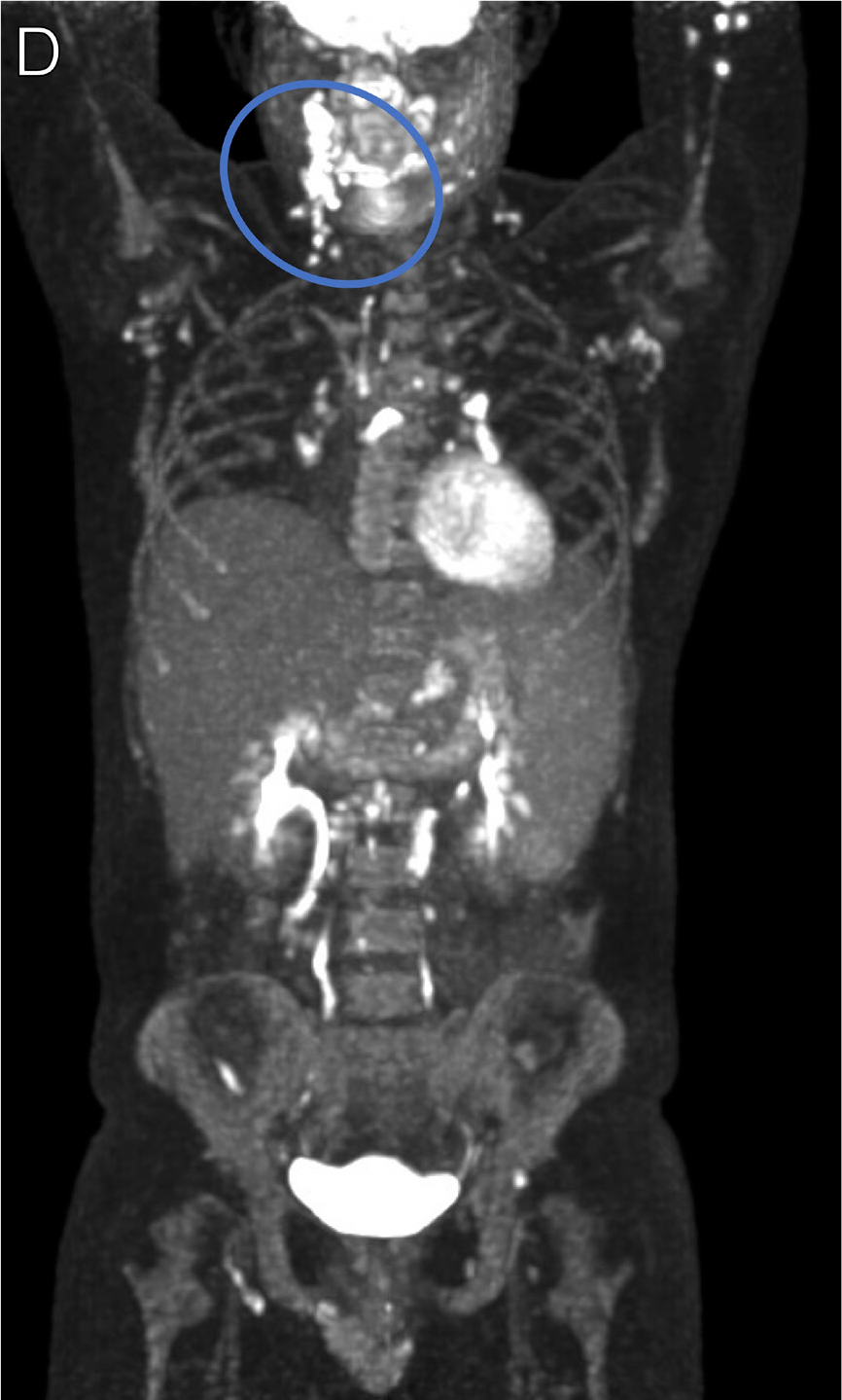
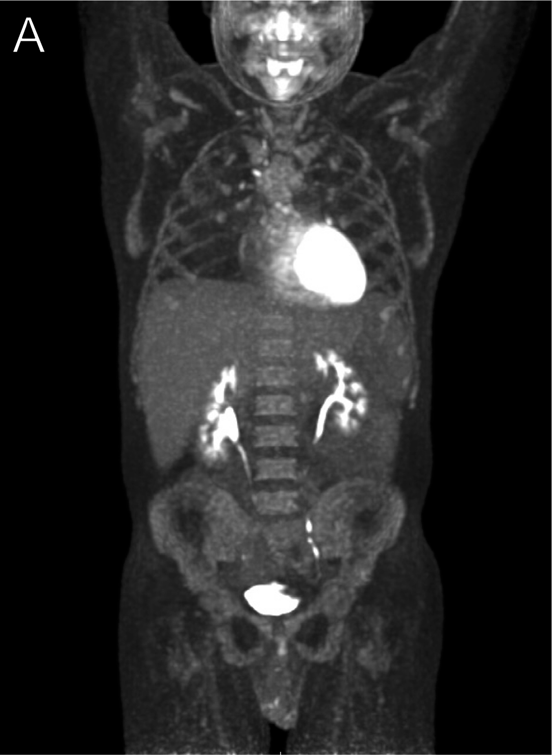
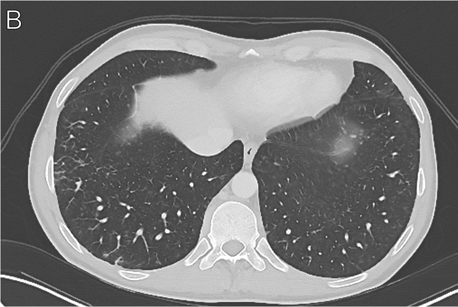
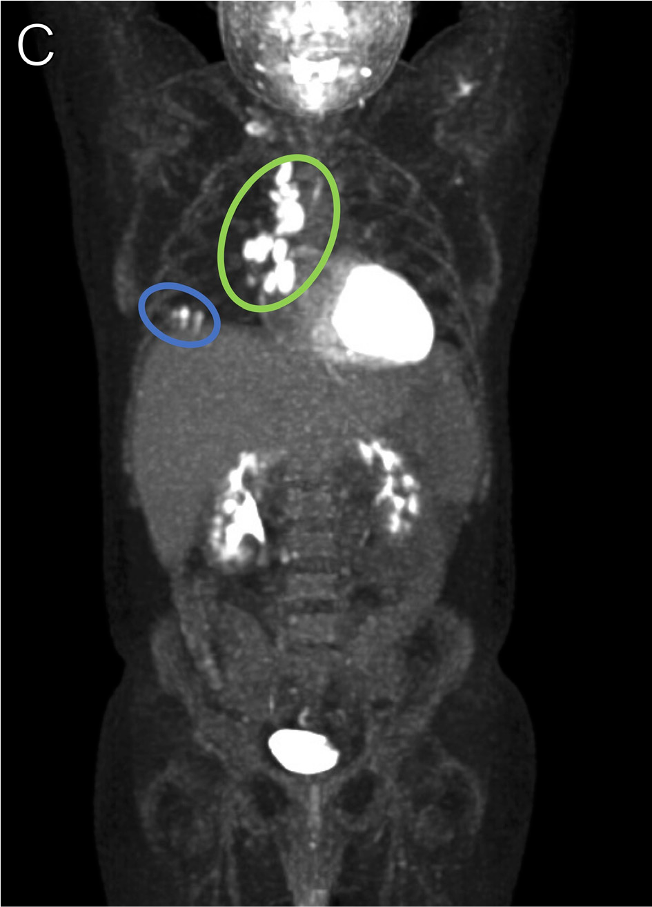
A man in his mid-30s originally from El Salvador presented with fevers, night sweats, weight loss, odynophagia, dyspnea, cough, and diarrhea. He was diagnosed with HIV, esophageal candidiasis, and histoplasmosis. Baseline PET-CT and CT scans of the chest are shown (Figures 19A-B). Six weeks after initiation of itraconazole and 4 weeks after ART initiation, he developed right mediastinal lymphadenopathy (Figure 19C, green circle) and right lower lobe consolidation (Figures 19C-D, blue circles) along with persistence of cough. Bronchoscopy was performed with negative cultures; however, cytopathology revealed GMS stain-positive organisms with morphology consistent with Histoplasma spp. Presentation was thought to be consistent with IRIS secondary to disseminated histoplasmosis.15,16
Human Herpesvirus-8 Disease
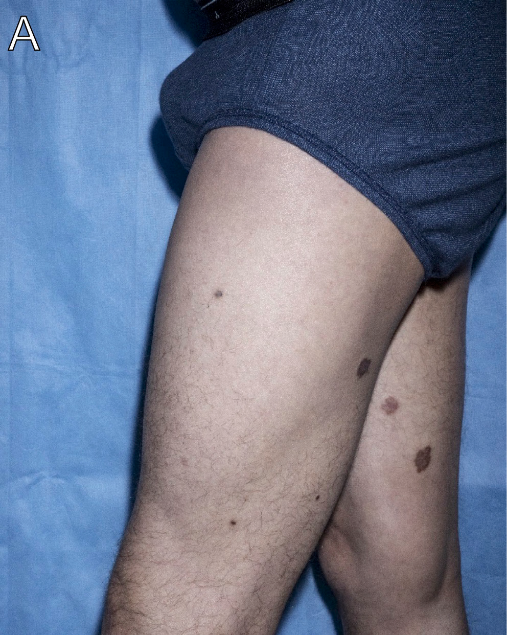
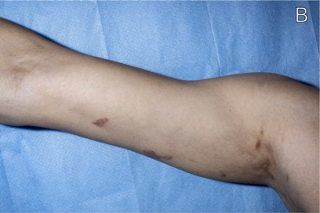
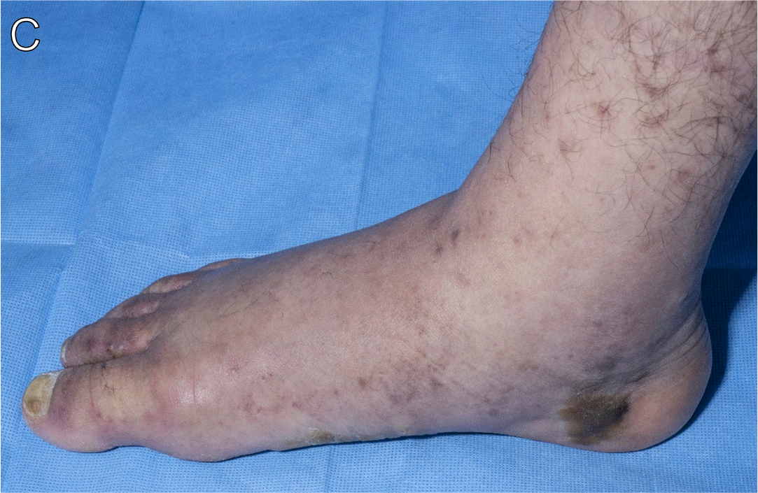
A man in his early 40s with HIV (CD4+ count, 18 cells/μL) not on ART presented with development of new skin lesions over the preceding year that were scattered over his chest, back, lower (Figure 20A) and upper (Figure 20B) extremities, torso, and feet (Figures 20C-D), as well as with a 1-week history of lower-extremity edema (Figure 20C). Biopsy of a representative lesion showed human herpesvirus-8 (HHV-8)-positive immunostaining in the vascular endothelial cells, consistent with Kaposi sarcoma (KS).17,18
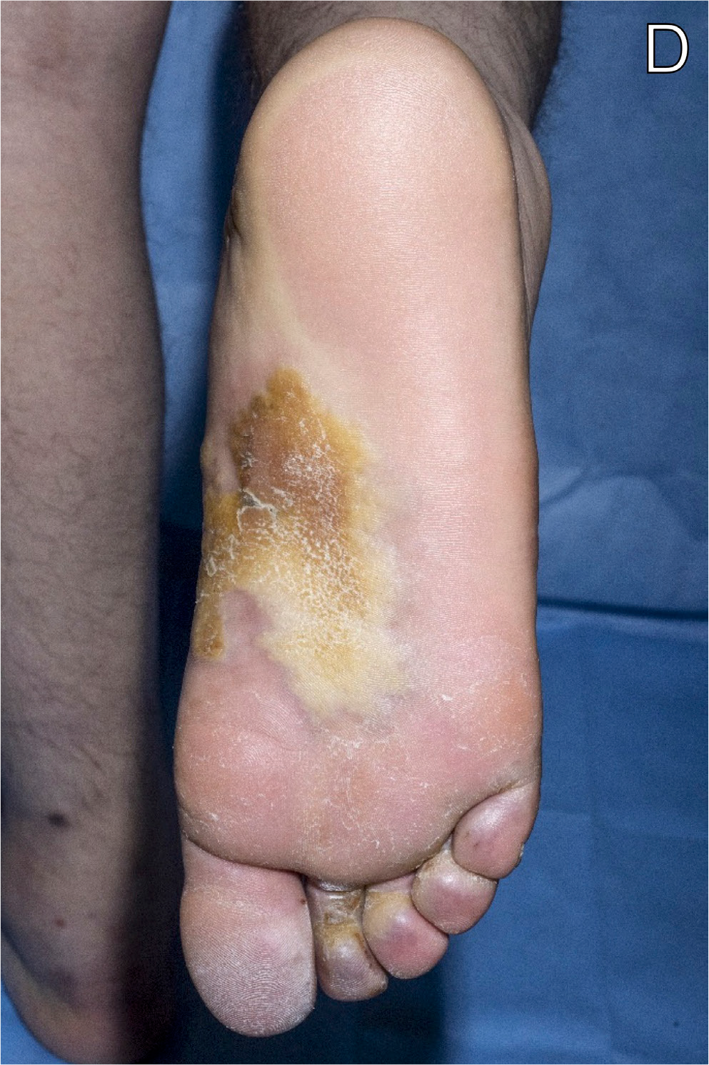
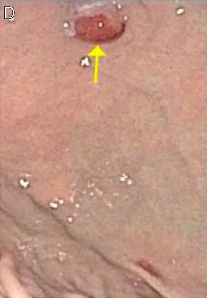
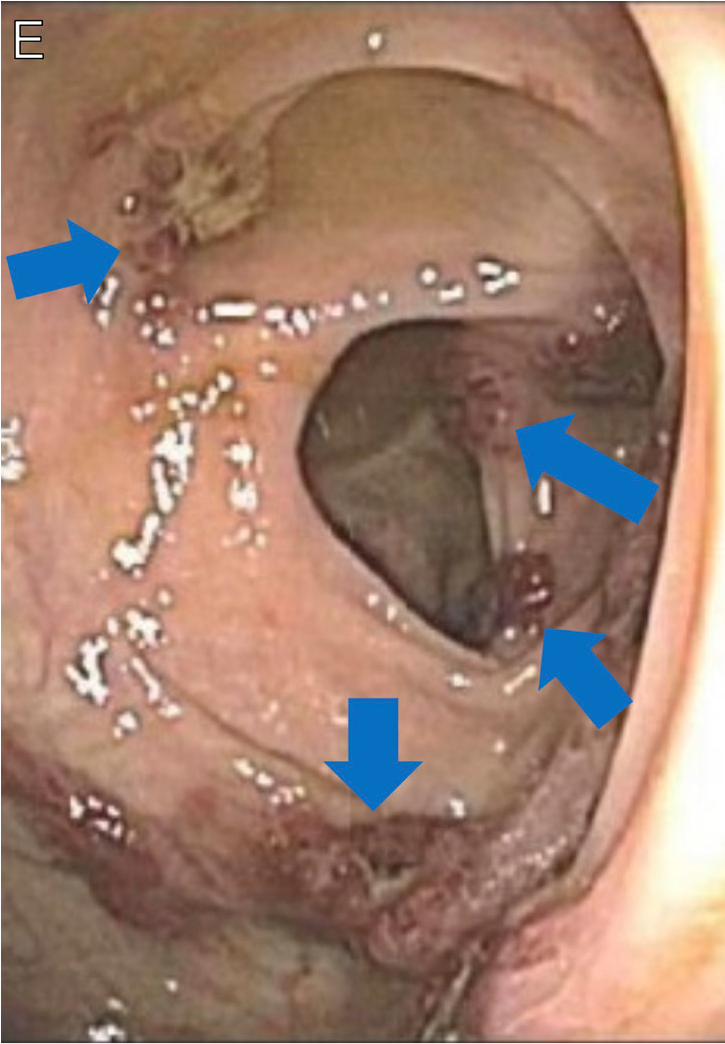
A man in his late 20s with HIV (CD4+ count, 224 cells/μL) presented after developing violaceous cutaneous patches (Figures 21A-C) 8 months after starting ART. Skin biopsy of a representative lesion was consistent with KS. Upper (Figure 21D, arrow) and lower (Figure 21E, arrows) endoscopies demonstrated extensive visceral involvement. Presentation was consistent with unmasking HHV-8 IRIS. He initiated doxorubicin for rapidly progressing KS with visceral involvement and KS inflammatory cytokine syndrome (KICS),17,18 and completed 7 cycles with clinical improvement.
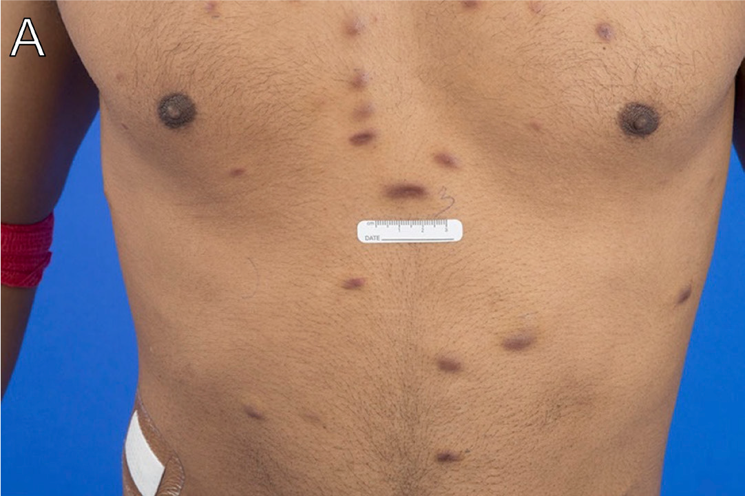
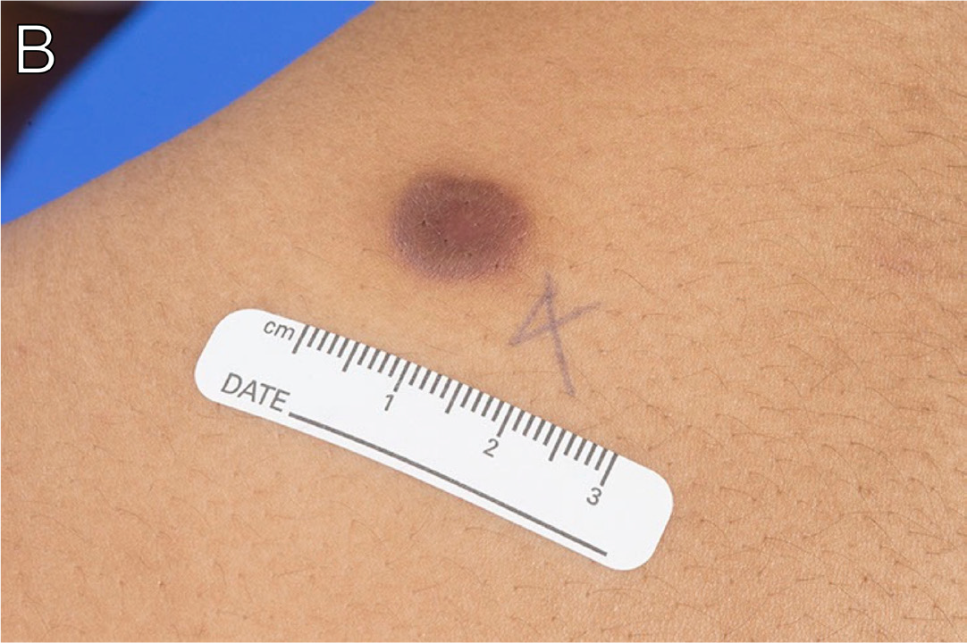
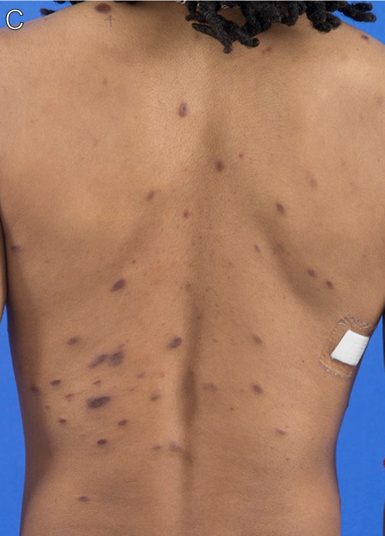
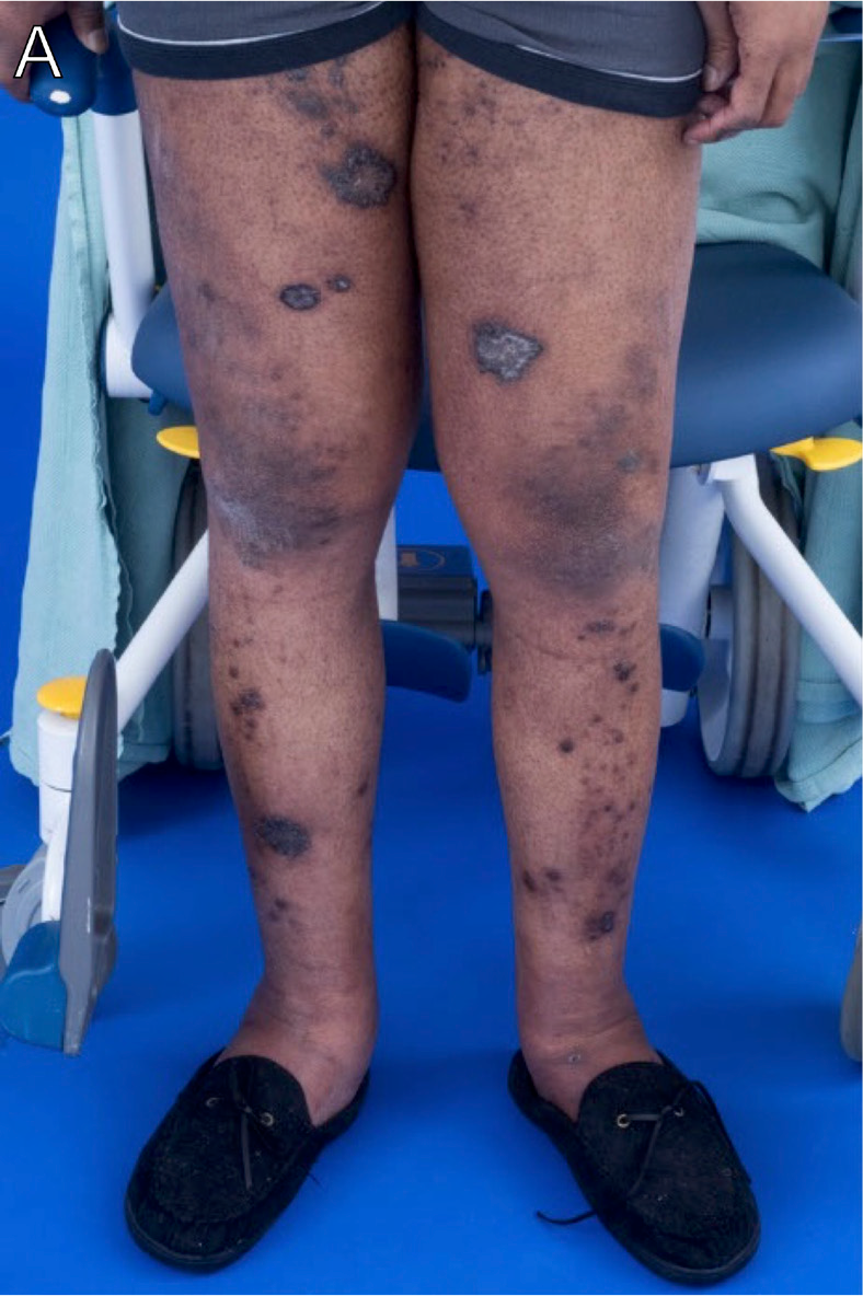
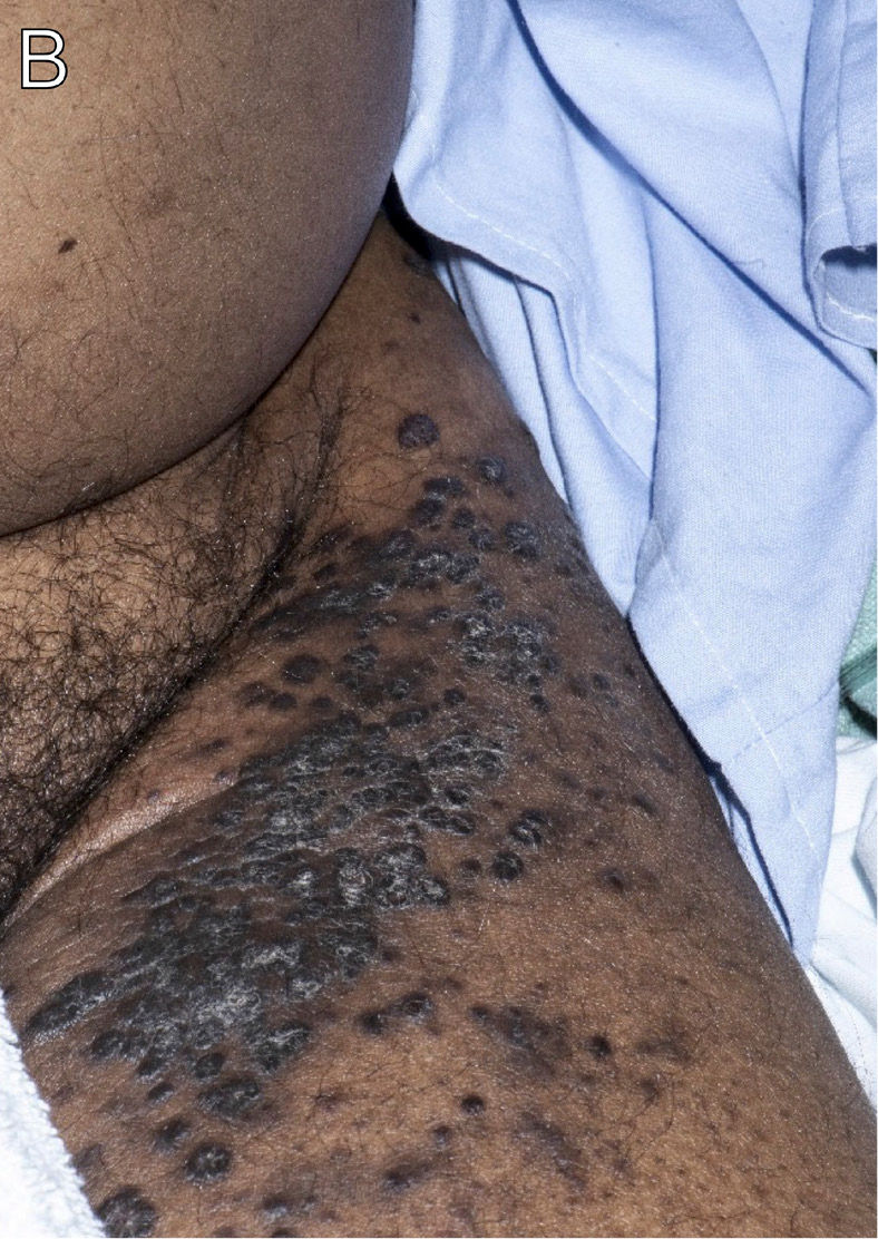
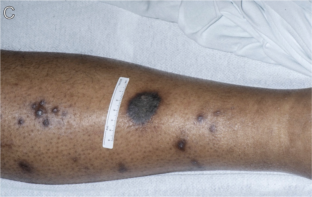
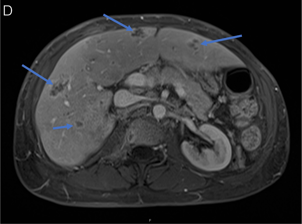
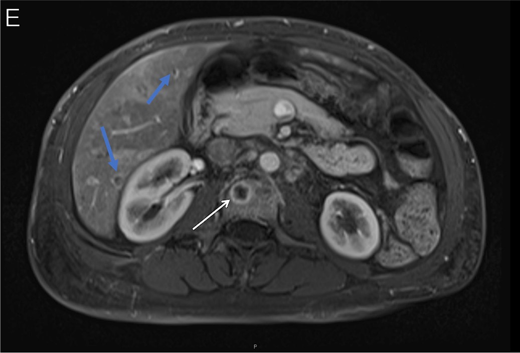
A man in his early 50s with HIV (CD4+ count, 4 cells/μL) not on ART presented with dyspnea, lower extremity edema (Figure 22A), and widespread violaceous cutaneous nodules (Figures 22A-C). Biopsy of a representative skin lesion was consistent with KS. On MRI of the abdomen, he was found to have numerous irregular lesions with peripheral enhancement distributed throughout the right and left lobes of the liver (Figures 22D-E, blue arrows). MRI also demonstrated heterogeneous bone marrow signal intensity with areas of irregular enhancement (Figure 22E, white arrow). He began treatment with ART and doxorubicin, but his treatment course was complicated by KICS, requiring initiation of methylprednisolone and rituximab.17,18
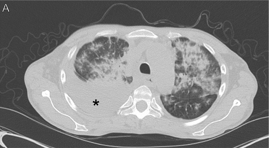
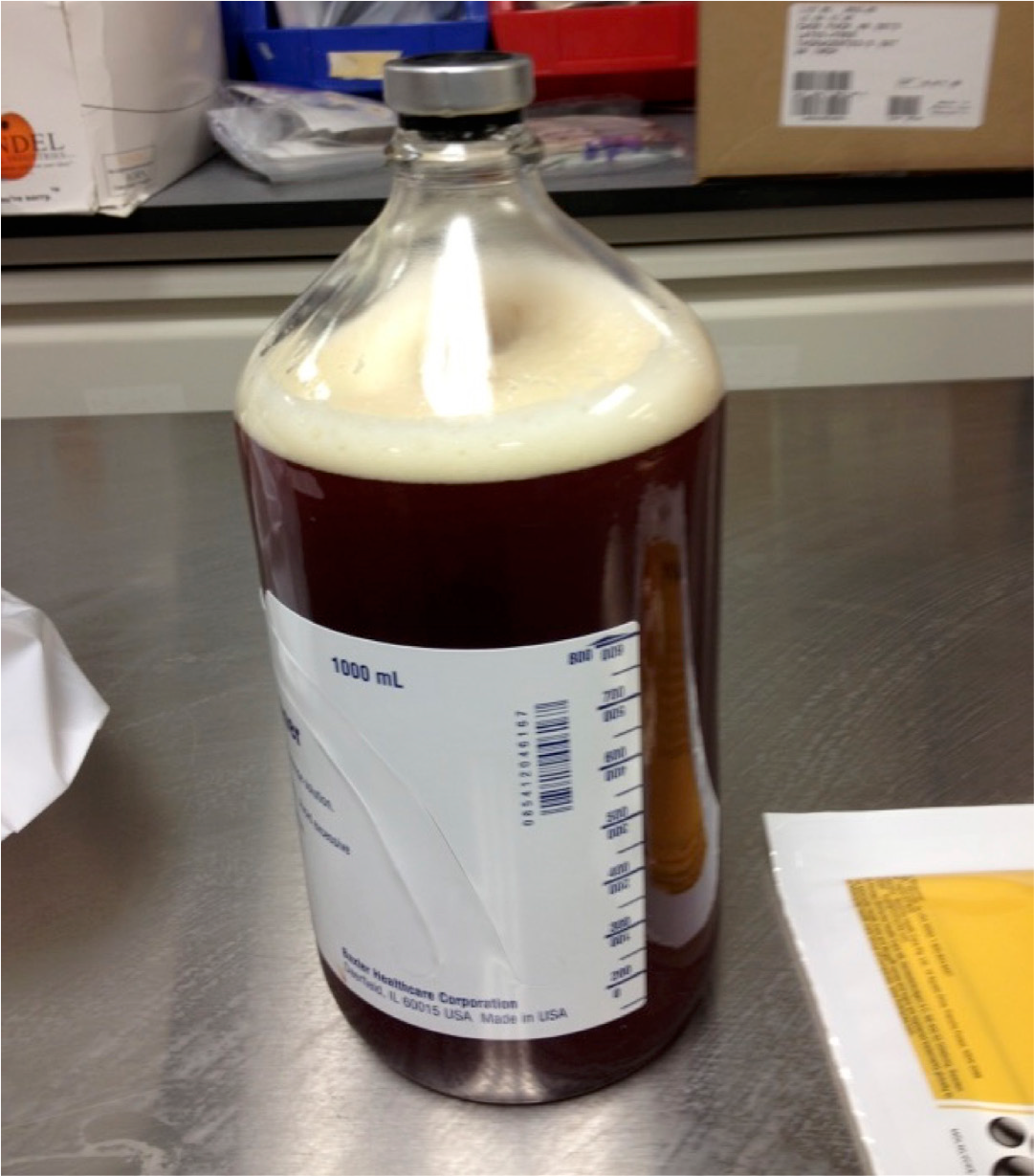
A man in his late 40s with HIV (CD4+ count, 49 cells/μL) not on ART presented with weight loss, dyspnea on exertion, cough, and skin lesions. A skin biopsy revealed KS. A CT scan of the chest demonstrated bilateral pulmonary infiltrates and right pleural effusion (Figure 23A, asterisk). A bronchoscopy was performed and showed hyperemic lesions (Figure 23B, arrows), consistent with visceral KS. A thoracentesis was completed and drained brown fluid (Figure 23C). Pleural fluid cytopathology showed scattered atypical lymphoid cells with enlarged, irregular nuclei, and a polymorphous population of lymphocytes. Immunocytochemical studies showed that the atypical lymphoid cells stained positive with antibodies to HHV-8, CD138, and CD38. In situ hybridization to EBER was positive in atypical cells. Presentation was consistent with primary effusion lymphoma (PEL). He initiated ART and began treatment for KS and PEL with rituximab, etoposide, prednisone, vincristine, cyclophosphamide, doxorubicin, and intrathecal methotrexate. Complete remission was achieved, but he developed recurrent PEL 12 months later. He subsequently received ifosfamide, carboplatin, etoposide, and lenalidomide, and complete remission was achieved.17,18
Human Papillomavirus
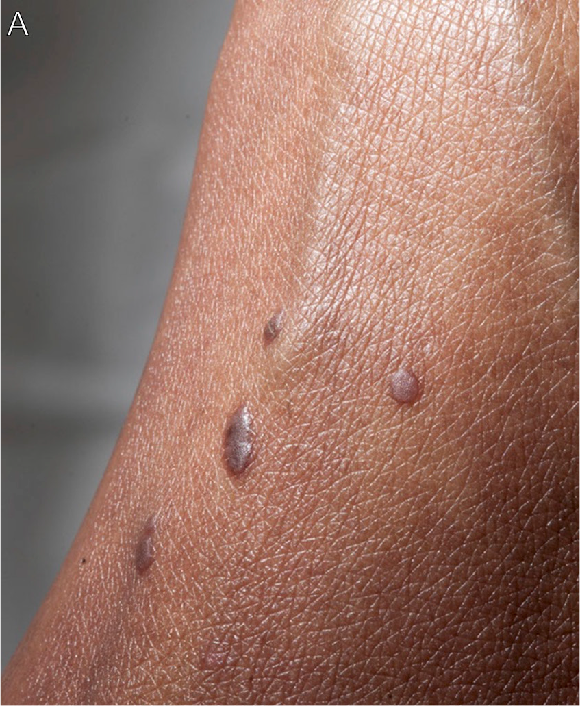
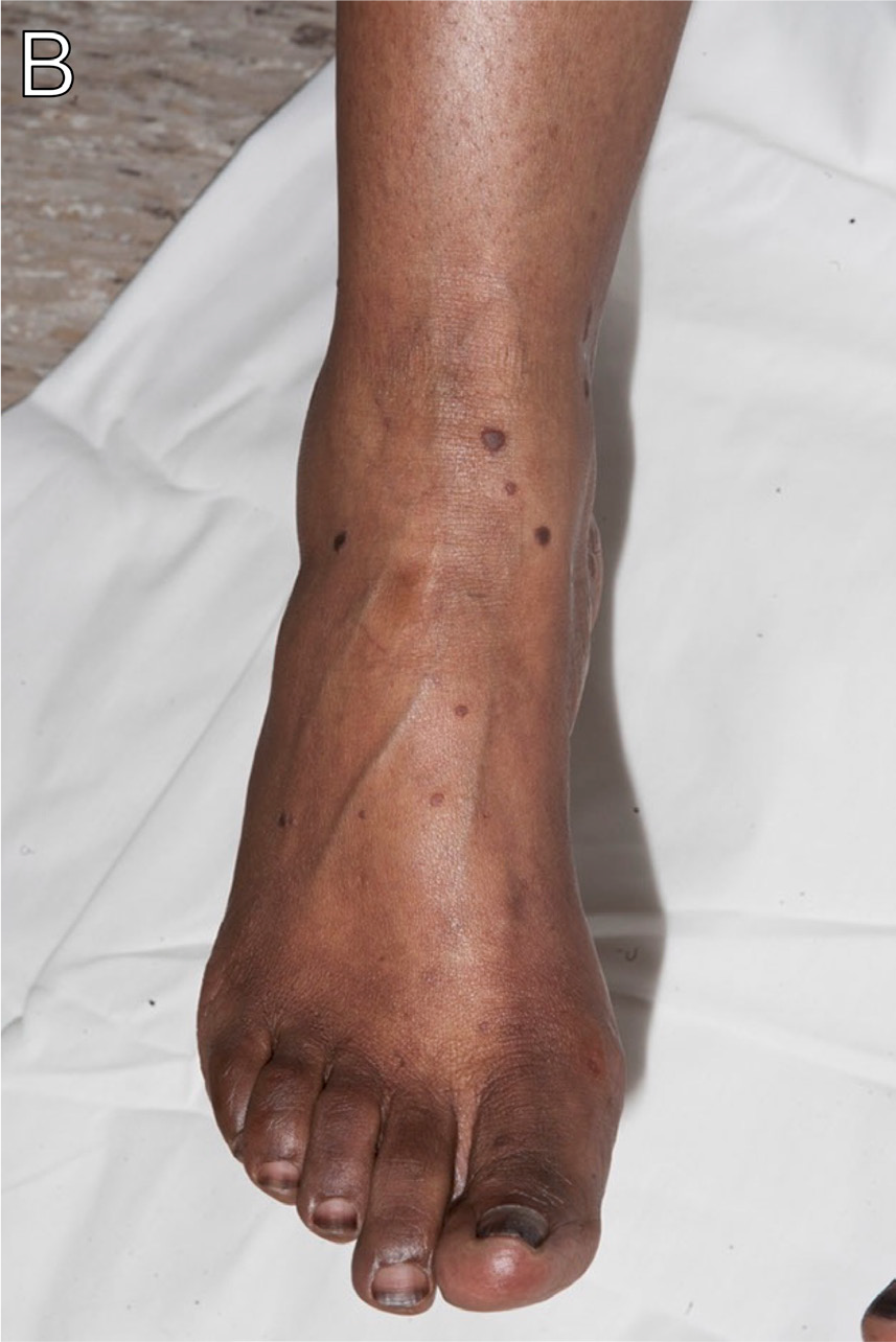
A woman in her late 30s with HIV (CD4+ count, 43 cells/μL) and diffuse large B-cell lymphoma status post treatment with rituximab, etoposide, prednisone, oncovin, cyclophosphamide, and hydroxydaunorubicin (R-EPOCH) presented 6 months after ART initiation with new violaceous papules and nodules on her lower legs (Figures 24A-B) and labia. Biopsy of a representative lesion on the leg was positive for human papillomavirus (HPV) and negative for HHV-819 consistent with severe verrucae.
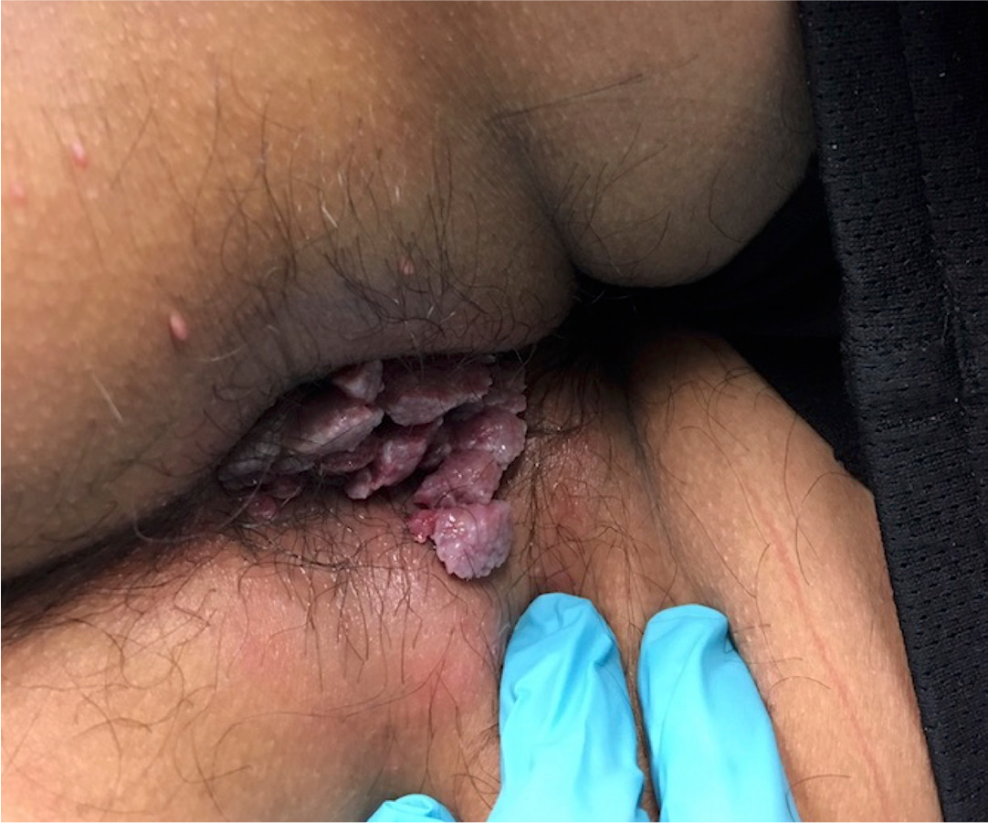
A man in his early 20s with HIV on ART presented with anal pruritus, discomfort with bowel movements, and blood with defecation for the preceding 2 weeks. Rectal exam revealed various verrucous lesions and inflamed erythematous tissue over the perirectal area (Figure 25). A herpes simplex virus (HSV)-2 PCR test was positive. He was diagnosed with condylomata acuminata and HSV proctitis. He initiated valacyclovir and topical imiquimod and was referred for colorectal surgery evaluation.19
Mycobacterial Infections
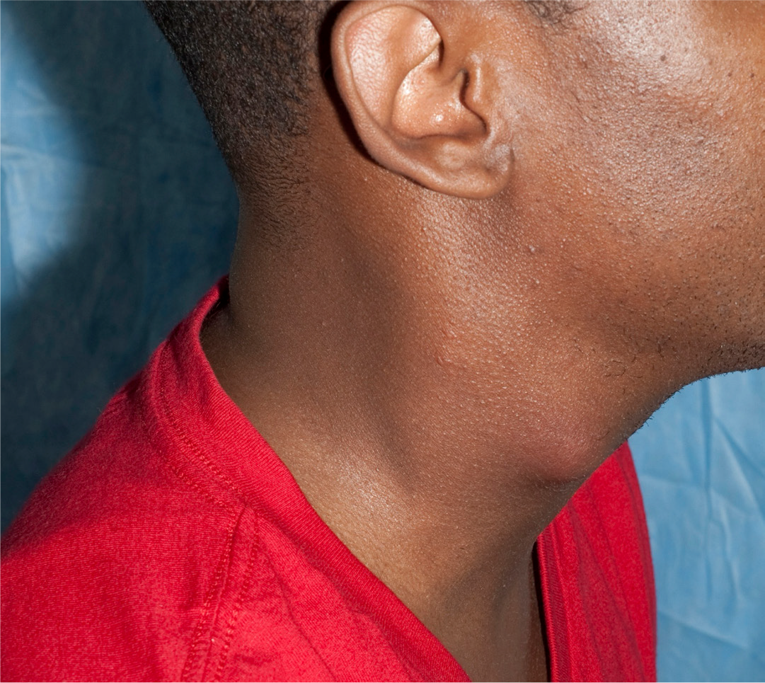
A man in his early 30s with advanced HIV developed a large lymph node on the right side of his neck (Figure 26) about 4 weeks after starting ART. Biopsy was performed and showed few acid-fast bacilli (AFB). Mycobacteria culture was positive for Mycobacterium avium complex (MAC), with presentation consistent with unmasking MAC IRIS.20
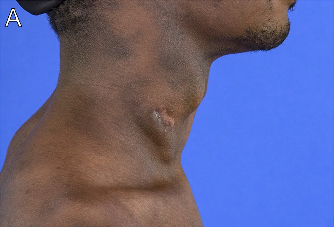
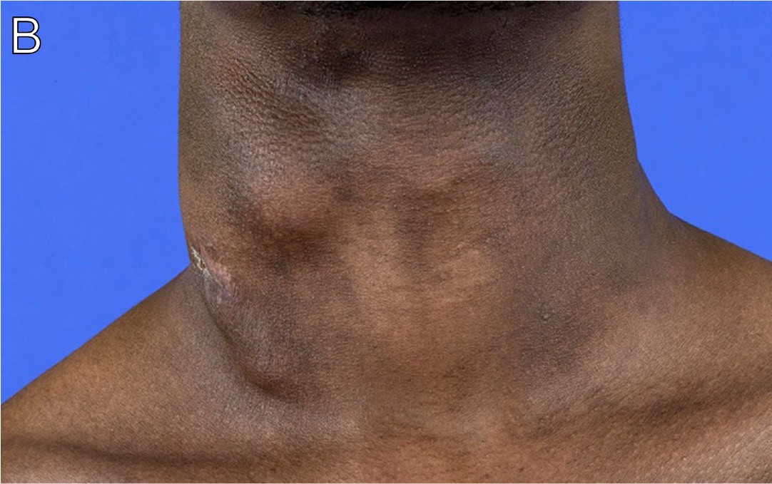
A man in his early 20s was diagnosed with HIV after presenting with oral thrush. Approximately 5 months after initiation of ART, he presented with enlarging right-sided cervical adenopathy. He had a nondiagnostic needle aspiration 10 months later. He underwent excisional biopsy of a lymph node 1 month later. Pathology demonstrated granulomas with reportedly negative infectious stains. He started empiric treatment with rifampicin, isoniazid, pyrazinamide, and ethambutol therapy for TB. His lymphadenopathy persisted, and he was referred to our institution. On exam, he had prominent right-sided cervical adenopathy (Figures 27A-B) that was hypermetabolic on PET-CT imaging (Figures 27C-D). He underwent repeat right cervical lymph node biopsy, which returned purulent fluid. Staining showed many AFB, and sequencing was consistent with MAC. His treatment was modified to azithromycin, ethambutol, and moxifloxacin.20 He had gradual improvement in the size of the lymphadenopathy with decreased metabolic activity seen on repeat PET scan after 2 years of therapy.
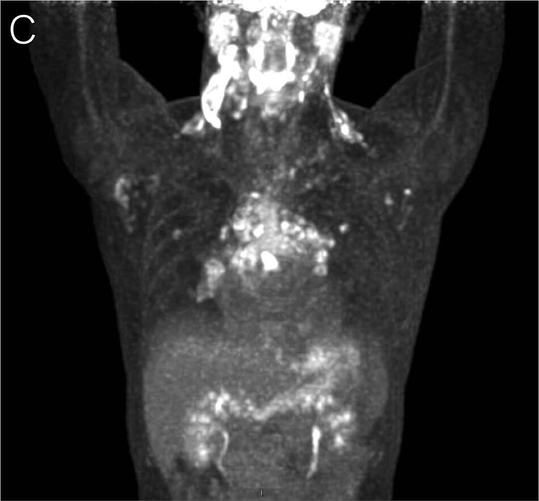
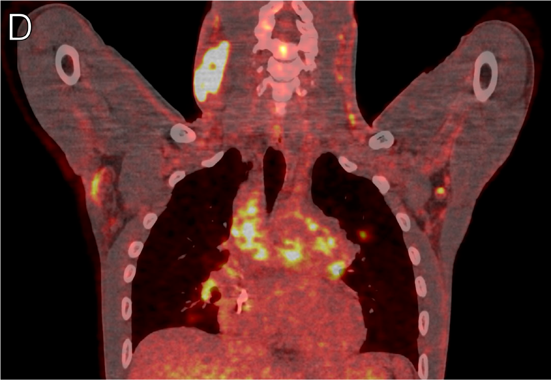
A man in his early 40s originally from Guatemala with HIV not on ART presented to care with a 4-month history of progressive weight loss, fatigue, dysphagia, intermittent fever, and progressive dyspnea. He was admitted to care with thrush, diffuse lung infiltrates on CT, anemia, and thrombocytopenia. He was diagnosed with Pneumocystis pneumonia and histoplasmosis, and started on ART. He presented to our institution with a CD4+ count of 3 cells/μL. CT imaging of the chest prior to ART revealed bilateral hilar and mediastinal lymph node enlargement (Figure 28A). Four weeks after ART initiation, he developed a new fever, and repeat CT imaging of the chest demonstrated increasing hilar and mediastinal lymphadenopathy (Figure 28B, arrow: confluent hilar lymphadenopathy measuring 3.7 × 2.4 cm). PET-CT imaging demonstrated nodular consolidations and increased mesenteric adenopathy (Figure 28C). Sputum cultures grew Mycobacterium avium, with presentation consistent with unmasking MAC IRIS.
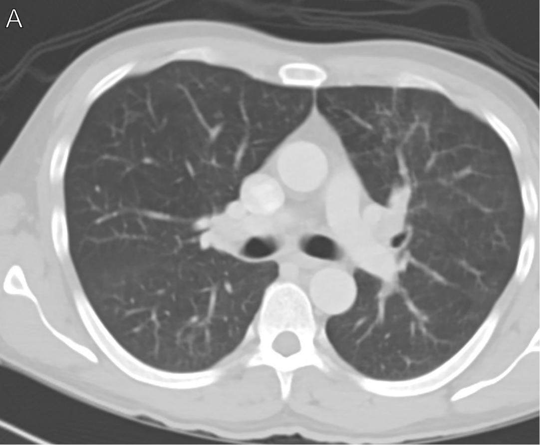
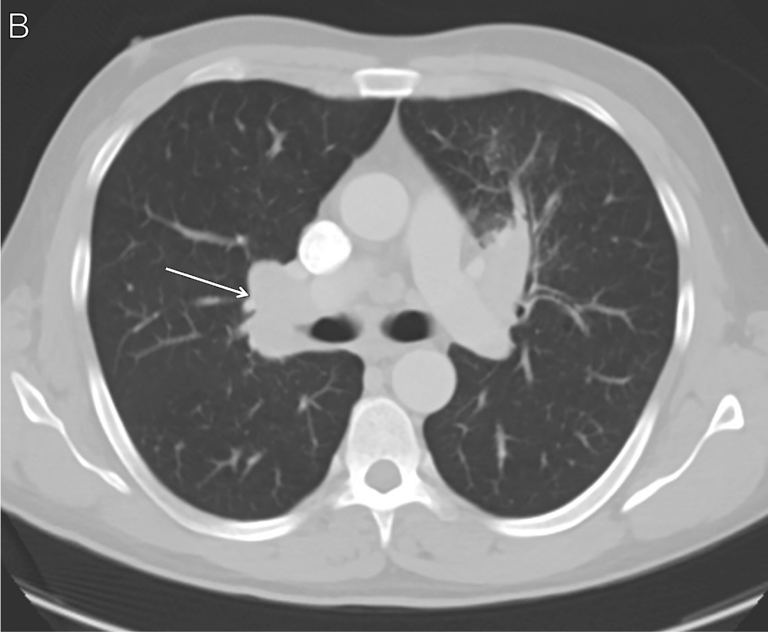
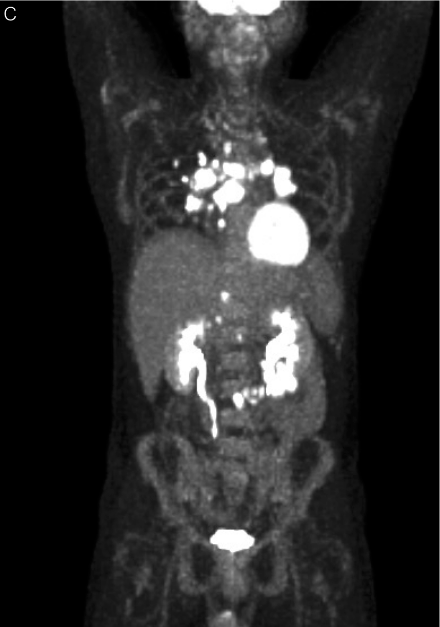
A man in his late 20s originally from Mexico presented with weight loss, abdominal pain, and neck swelling. CT imaging revealed cervical and supraclavicular adenopathy. He underwent a biopsy that was nondiagnostic. Two months later he re-presented with a 20-pound weight loss, fever, nausea, vomiting, and diarrhea. CT imaging revealed pleural effusions and ascites. He was diagnosed with HIV and had a CD4+ count of 21 cells/µL. A cervical lymph node biopsy revealed rare AFB on stain and was culture positive for Mycobacterium tuberculosis complex. He was started on TB therapy and ART. He re-presented approximately 1 week later with dyspnea and pleuritic chest pain, was diagnosed with bilateral pulmonary emboli, and was started on anticoagulation. However, he had persistent fevers, recrudescence of abdominal pain, and swelling along with dysuria. He was referred to our institution for further evaluation. PET-CT imaging was notable for left cervical, axillary, and mediastinal adenopathy as well as peritoneal and numerous abdominal and pelvic areas of hypermetabolic activity (Figures 29A-B). Genitourinary involvement was found, with positive urine AFB stain and hypodensities on the kidneys (Figure 29C, arrow). His antimycobacterial regimen was intensified with the addition of moxifloxacin and linezolid, and corticosteroids were initiated. His symptoms improved. With the extensive peritoneal and genitourinary involvement, it was suspected the patient had Mycobacterium bovis, which was confirmed on sequencing of the patient's initial lymph node biopsy culture.21
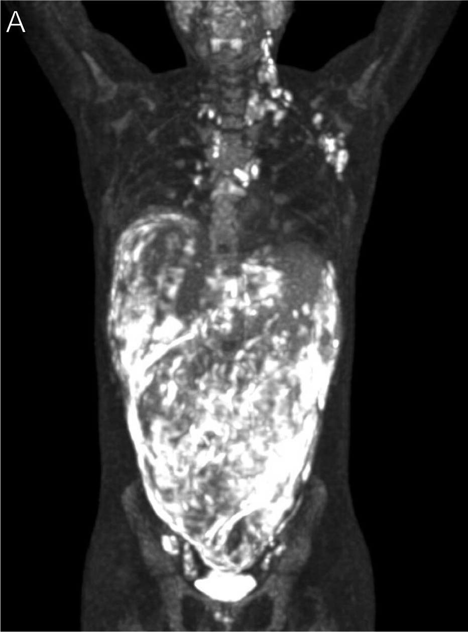
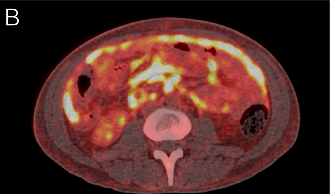
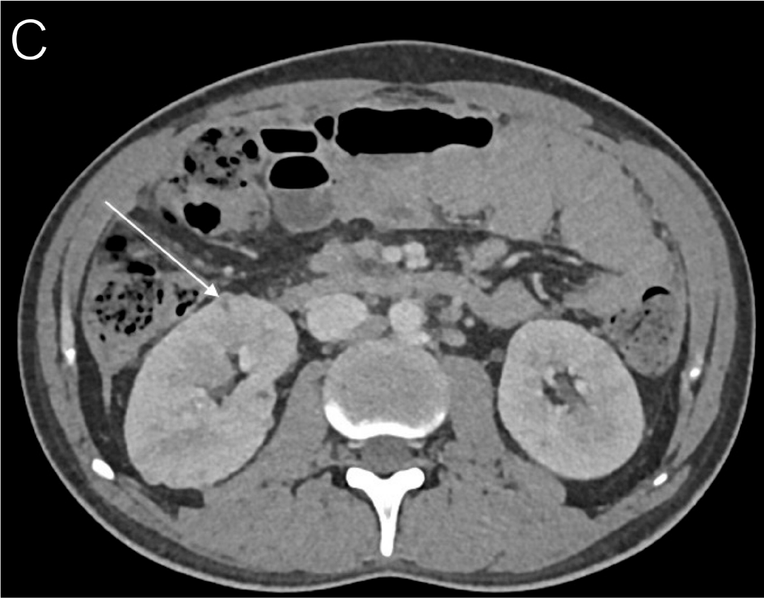
A woman in her late 30s originally from Cameroon presented with progressive fatigue, weight loss, respiratory symptoms, and fevers for 4 months. CT imaging demonstrated a miliary pattern in the lungs, bronchiectasis, and lung nodules. Her sputum cultures were AFB positive and later grew Mycobacterium tuberculosis. An HIV test was positive, and she had a CD4+ count of 38 cells/µL. She initiated TB therapy, but her treatment course was complicated by respiratory failure requiring high-flow oxygen. She developed rising transaminase levels that improved with discontinuation of rifampicin. However, despite improvement, she continued to have persistent fevers and was transferred to our institution. PET-CT imaging was notable for disseminated disease with pulmonary and bone involvement, as well as hepatosplenomegaly (Figure 30A). A chest CT scan further delineated miliary involvement (Figure 30B), and brain MRI noted left-sided mastoiditis (Figure 30C). Biopsy of the left hip demonstrated necrotizing granulomas (Figure 30D) and AFB (Figure 30E). She began ART, but her course was complicated by recrudescent fevers consistent with TB IRIS, which improved with initiation of corticosteroids.22
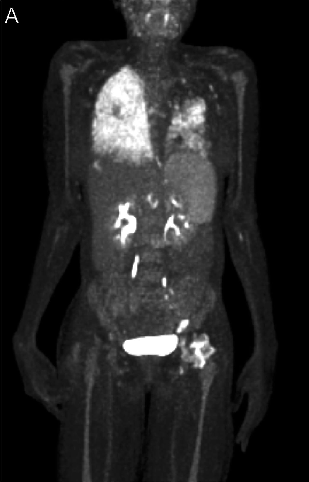
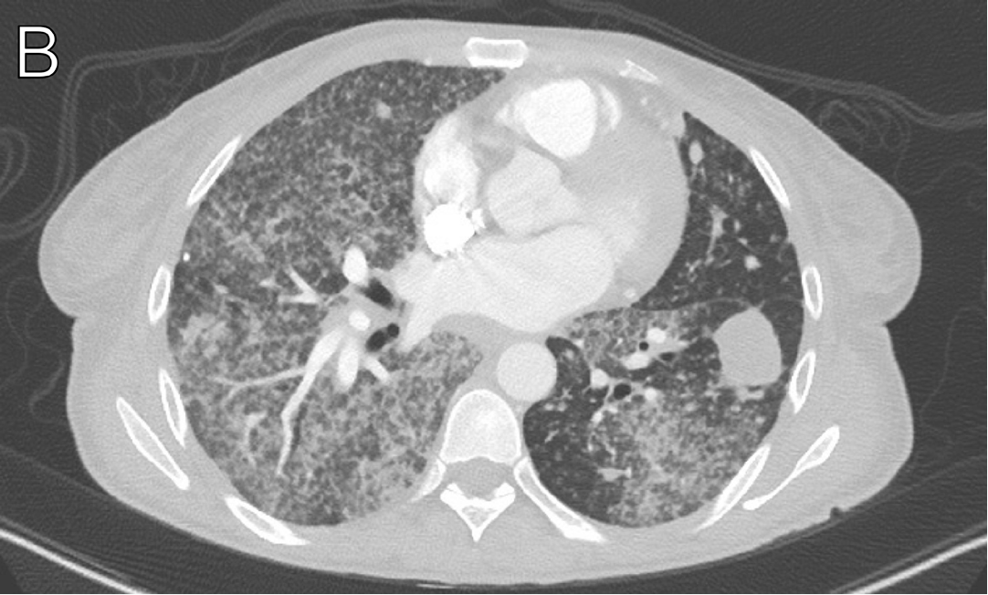
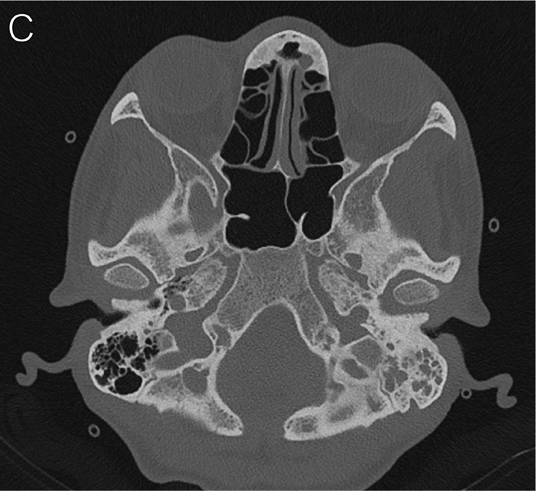
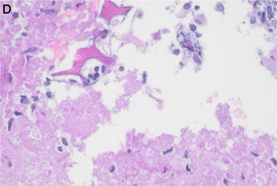
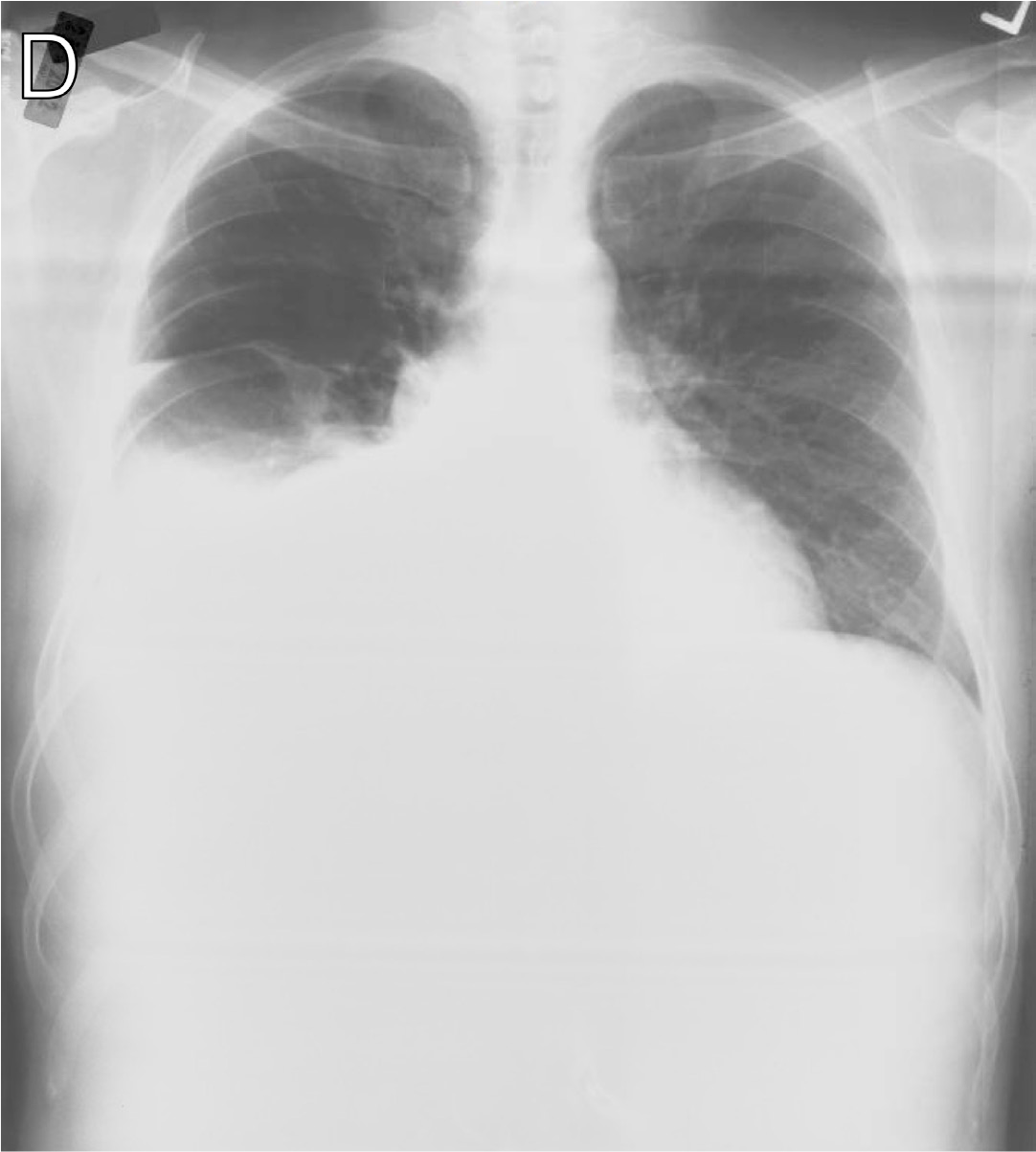
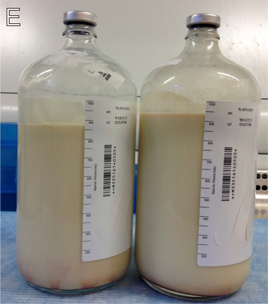
A man in his early 40s was diagnosed with HIV, right knee arthritis, tibial osteomyelitis (Figure 31A), and miliary pneumonia from drug-sensitive Mycobacterium tuberculosis (Figure 31B). TB medications and, subsequently, ART were initiated.22 Two weeks later, fever and knee pain recurred. His HIV RNA level was 747 copies/mL, and his CD4+ count was 171 cells/µL (from 105,751 copies/mL and 20 cells/µL, respectively). Repeat MRI showed increased enhancement and increased extraosseous component of the tibial infection (Figure 31C). Repeat synovial tissue biopsy was negative for mycobacteria. Prednisone was started for IRIS with clinical and radiologic improvement. Five weeks later, he developed shortness of breath and cough, and a chest X-ray revealed a new right pleural effusion (Figure 31D). Two liters of milky fluid were drained with thoracentesis (Figure 31E). Microbiologic work-up was negative. Short-term fat-free diet, octreotide, and 1 infusion of infliximab during a prednisone taper led to a complete recovery.23
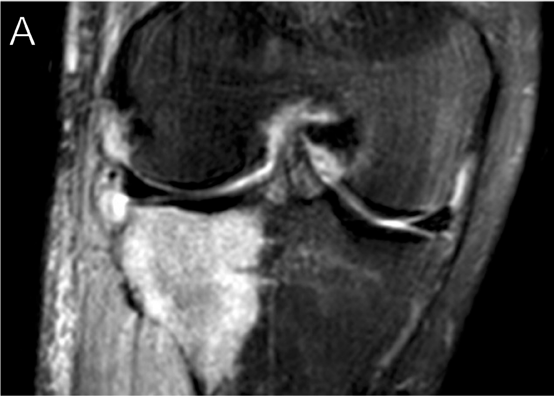
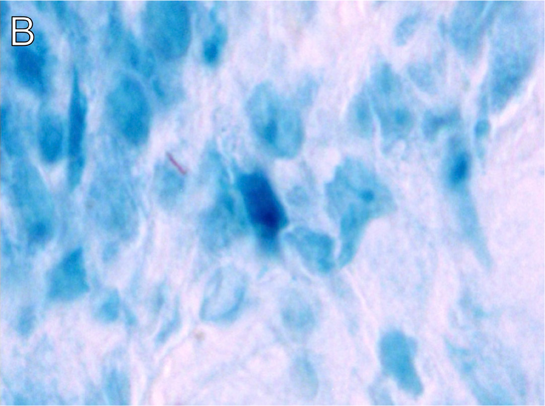
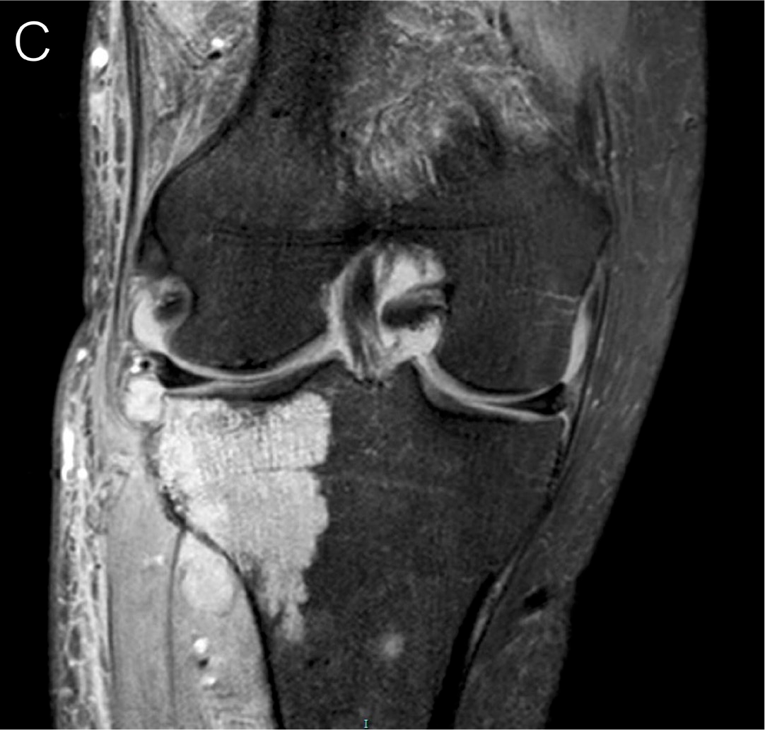
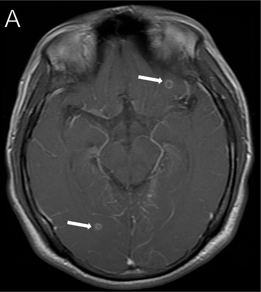
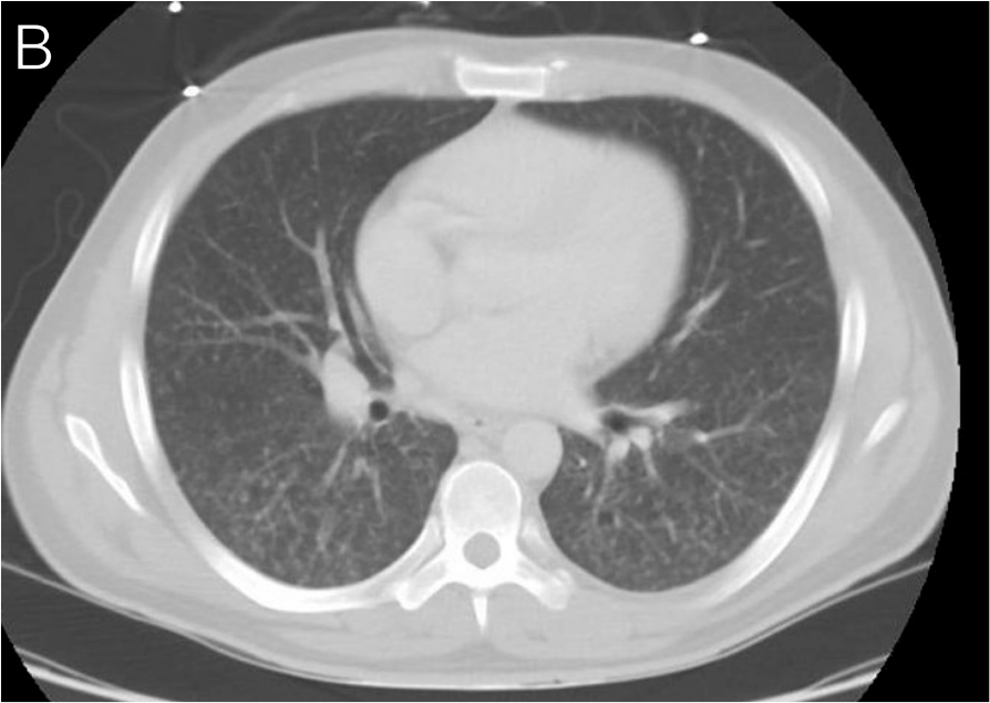
A man in his late 20s originally from Guatemala presented with a 2-week history of headaches, neck .pngfness, and visual deficits. On MRI of the brain, he was found to have ring-enhancing lesions (Figure 32A, arrows) along with pleocytosis in his CSF, and a CT scan of the chest demonstrated a miliary pattern (Figure 32B). He was diagnosed with HIV and TB, which was later confirmed as Mycobacterium bovis. He initiated TB therapy and corticosteroids with improvement in his symptoms and was referred to our institution. Approximately 4 weeks following initiation of TB therapy, his course was complicated by worsening recrudescent headaches and visual deficits. MRI was performed and showed interval progression of disease and meningitis concerning for paradoxical reaction (Figures 32C-D). The pattern of his visual field defects was notable for an incongruous right homonymous hemianopsia. His corticosteroids were increased with improvement of symptoms, and he underwent slow corticosteroid taper and subsequent ART initiation.22
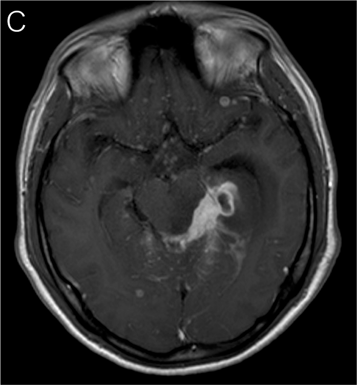
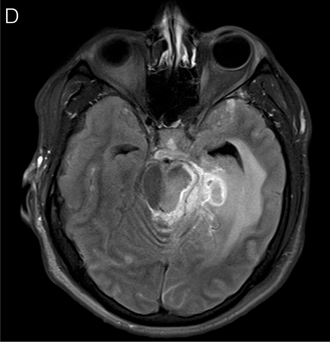
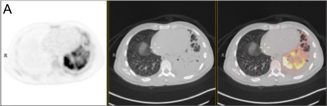
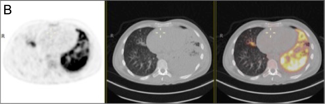
A man in his mid-30s originally from Guatemala presented with a 3-month history of cough, weakness, fatigue, weight loss, loss of appetite, and subjective fevers. He was diagnosed with pulmonary TB (Figure 33A) and HIV and referred to our institution. His CD4+ count was 29 cells/µL, and his HIV RNA level was more than 1 million copies/mL. He was initiated on TB therapy and had initial improvement of symptoms. Four weeks after initiation of TB therapy and on the day of ART initiation, he noted weakness in his right hand, with difficulty flexing his fourth and fifth digits along with numbness. He also noted a persistent cough. A repeat CT scan of the chest revealed enlargement of the left lung cavitary lesion, with PET-CT imaging demonstrating increasing hypermetabolic activity in this area (Figure 33B). MRI of the brain was performed and demonstrated focal leptomeningeal enhancement in the right temporal and occipital regions, with edema in the right occipital lobe and a 6-mm enhancing focus in the right cerebellar region, consistent with tuberculoma (Figure 33C). Microbiologic work-up by lumbar puncture was performed and no other infectious etiology was found. TB therapy was intensified, and corticosteroids were added, with which he improved.22
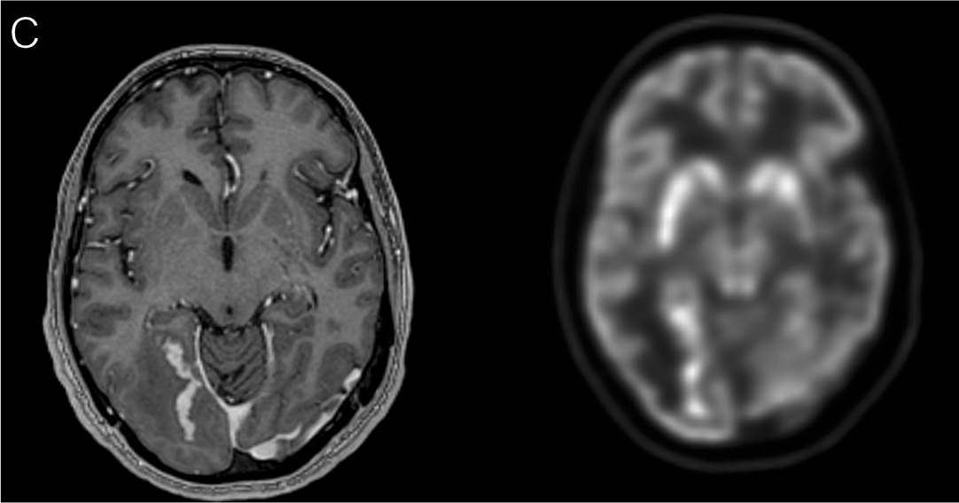
A woman in her mid-40s with HIV (CD4+ count, 28 cells/μL) not currently on ART presented with abdominal pain, distension, fevers, weight loss, and painful cutaneous nodules (Figure 34A). A CT scan showed substantial mesenteric lymphadenopathy. Lymph node biopsy revealed abundant AFB on Fite stain. 16S RNA sequencing returned positive for Mycobacterium genavense. Her clinical course was complicated by mycobacterial IRIS after she reinitiated ART. PET scan showed substantial uptake within the abdomen and numerous subcutaneous areas in her legs (Figure 34B, arrows). Upper endoscopy with push enteroscopy showed mucosal changes (Figure 34C) concerning for mycobacterial infection, with pathology revealing numerous AFB on Fite stain (Figure 34D). Acid-fast stain of the stool showed many AFB (Figure 34E). Her antimycobacterial regimen was intensified and she was treated with a prolonged corticosteroid taper, on which she improved.24
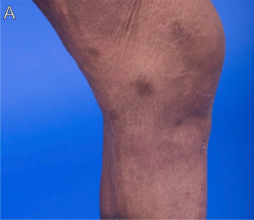
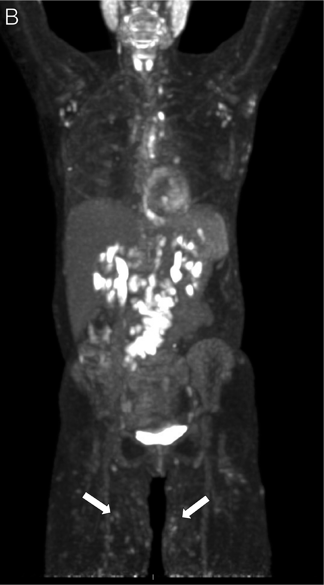
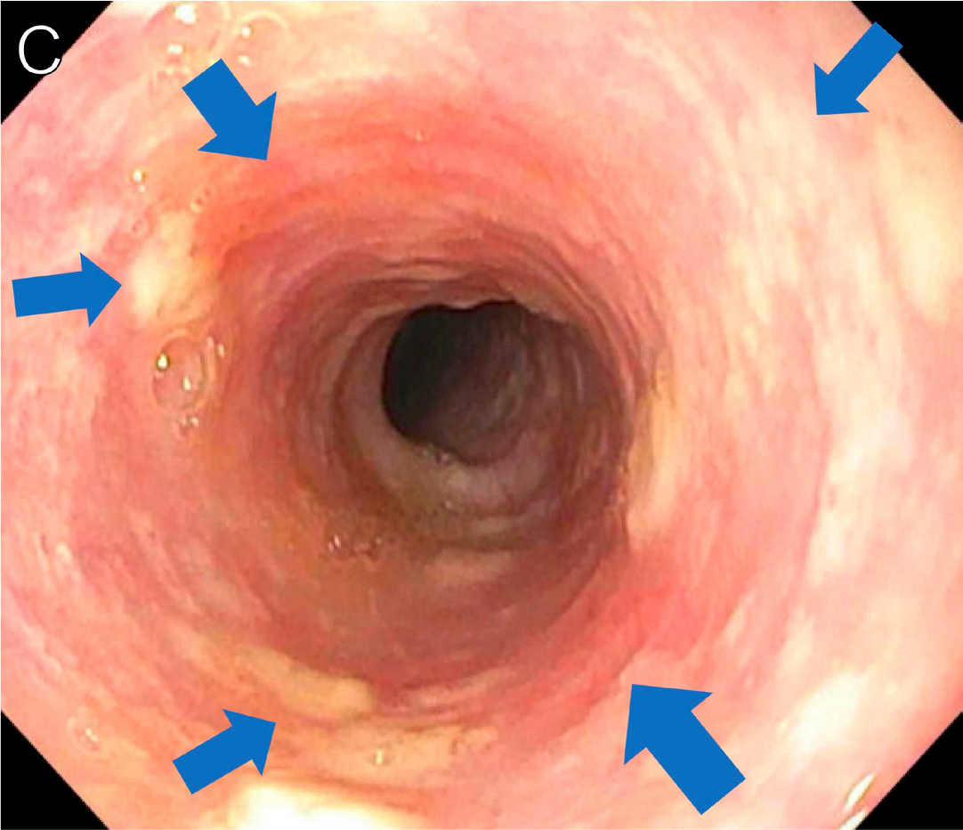
A man in his 20s presented 3 months after HIV diagnosis (CD4+ count, 7 cells/µL) and ART initiation with diarrhea, abdominal pain, skin nodules (Figures 35A-B, arrows), hepatosplenomegaly (Figure 35C), and lymphadenopathy. He was diagnosed with disseminated Mycobacterium genavense by PCR sequencing in skin, duodenal, and rectal biopsies. He was referred to our institution with persistent symptoms despite antimycobacterial therapy. Numerous biopsies from skin, bone marrow, and lymph nodes were obtained and demonstrated an overwhelming pathogen burden (Figure 35D). He had a protracted course but did slowly improve with intensification of his therapy with a course of interferon gamma therapy and additional antimycobacterial agents.24
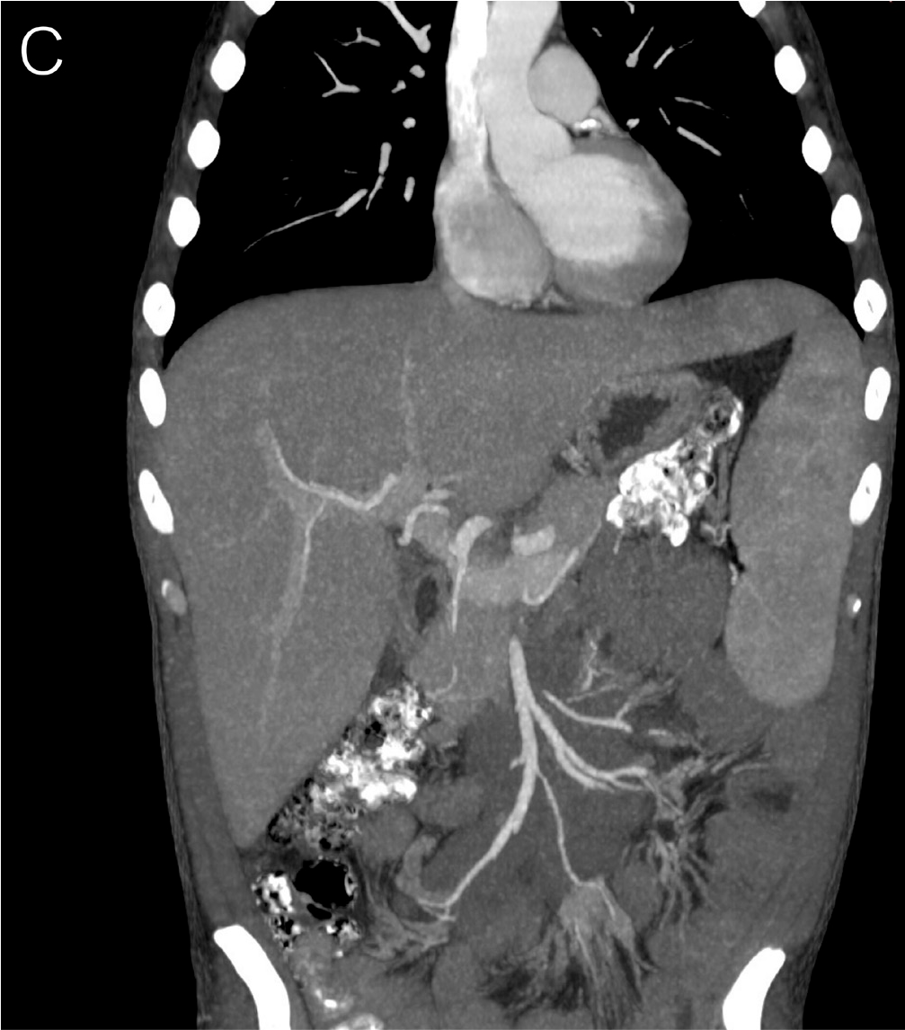
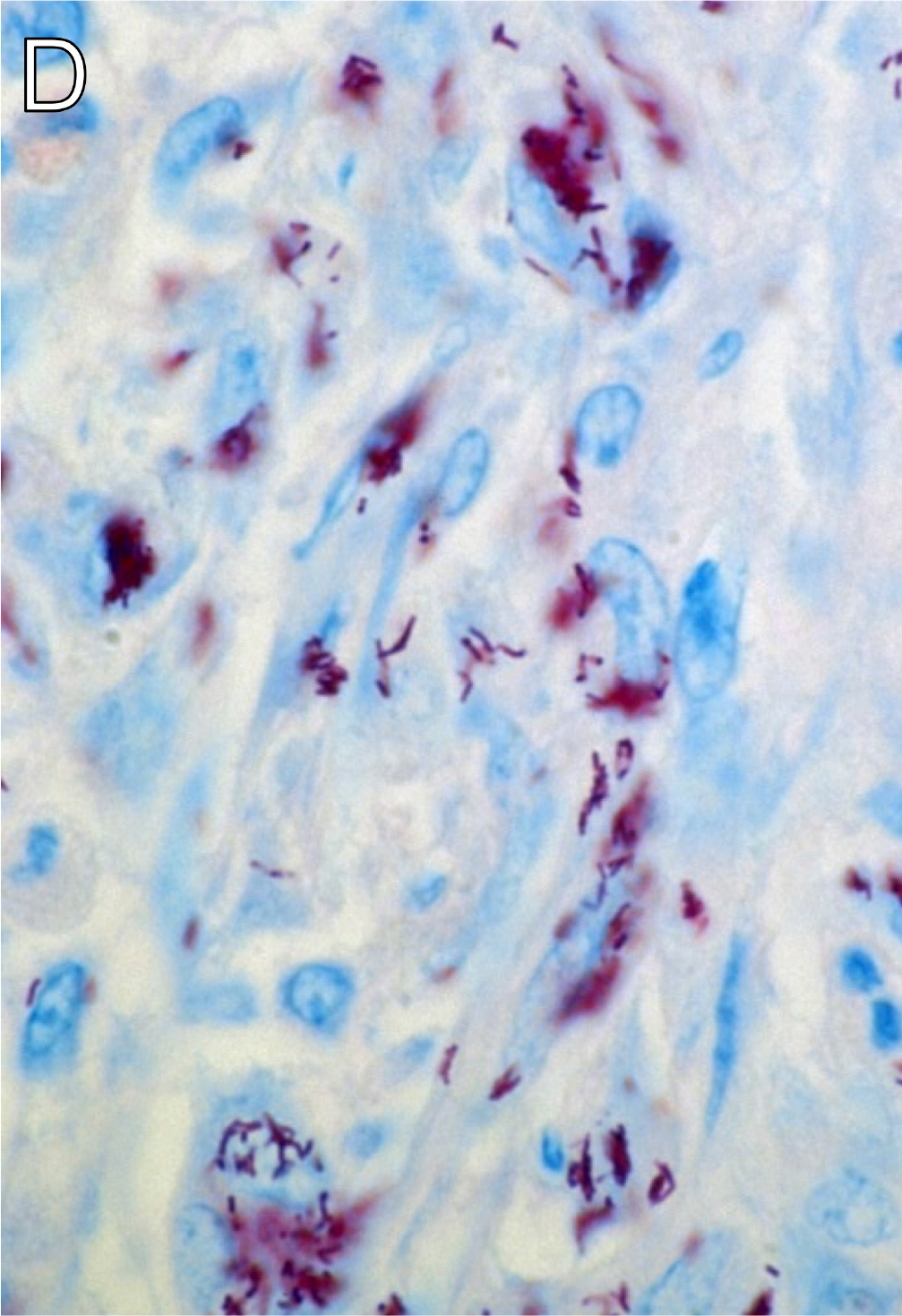
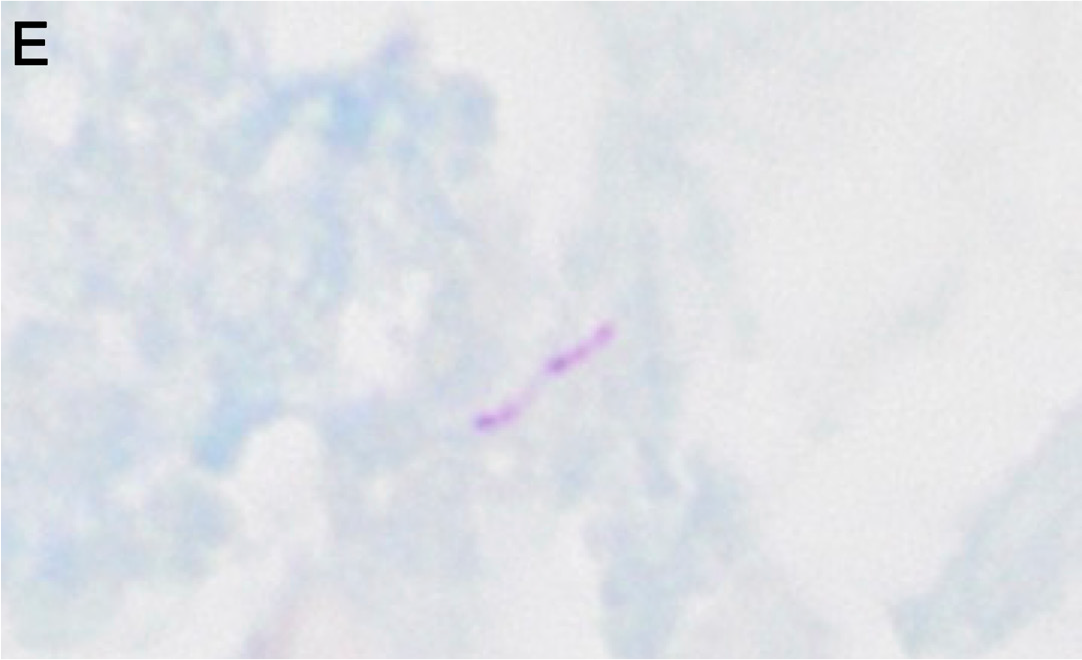
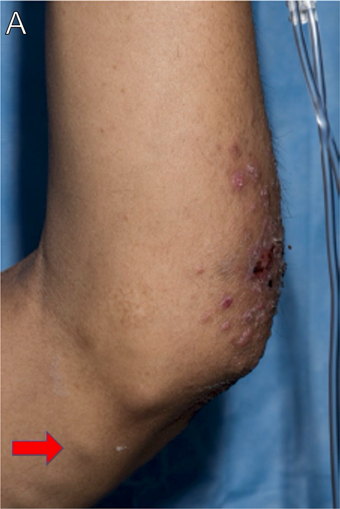
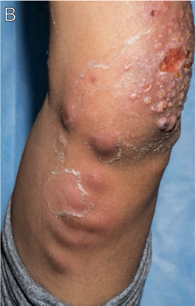
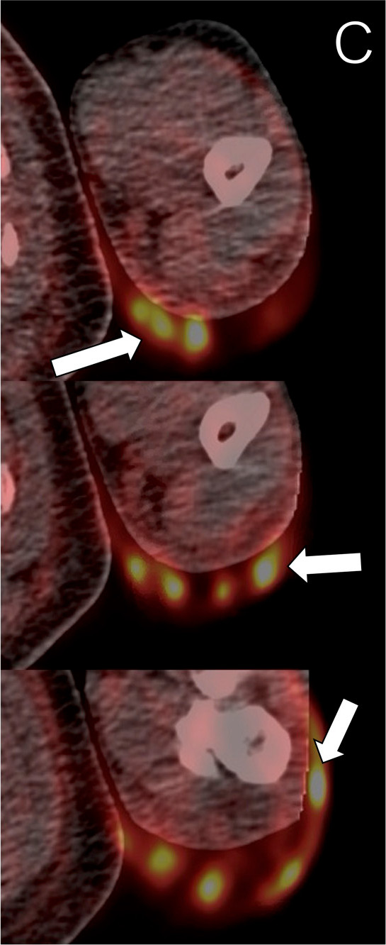
A man in his 30s who was a fish tank owner and worked as a pet groomer presented with a 4-month history of fatigue and oral and nasal ulcerations; a 2- to 3-month history of nodular lesions on his left elbow; and a 1-month history of fevers, chills, fatigue, diarrhea, and weight loss. When he presented to care, he was diagnosed with HIV and disseminated histoplasmosis. He was also found to have a nonhealing wound over his left elbow with nodular lymphangitis (Figure 36A, arrow; Figure 36B). His nonhealing wounds over his left elbow were biopsied and were AFB positive on pathology with sequencing of the culture revealing Mycobacterium marinum. He was started on antimycobacterial therapy and ART. Days after starting ART, his nodular lymphangitis over his left extremity became erythematous, edematous, and painful. The lymphadenopathy was found to be hypermetabolic on PET-CT imaging (Figure 36C, arrows). Given the proximity to ART initiation, the presentation was thought to be consistent with mycobacterial IRIS and improved with initiation of corticosteroids.25
Pneumocystis Pneumonia
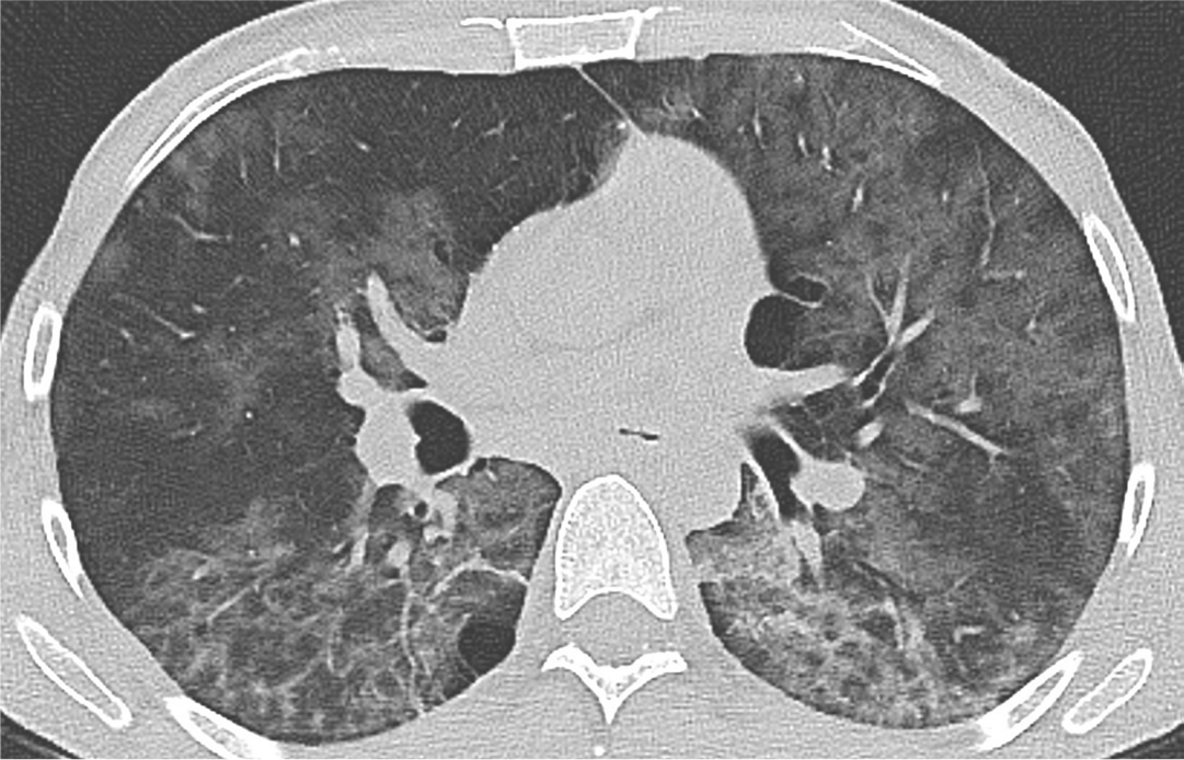
A man in his late 30s with newly diagnosed HIV (CD4+ count, 13 cells/µL) not on ART reported dyspnea on exertion and cough for the previous 2 days. CT imaging of the chest in lung windows demonstrated lobular areas of ground-glass attenuation, predominantly within the upper lobes (Figure 37). Patchy, peribronchovascular ground-glass opacities were present in the lower lobes. Pneumocystis direct fluorescent antibody (DFA) staining on a sputum sample was positive.26–28
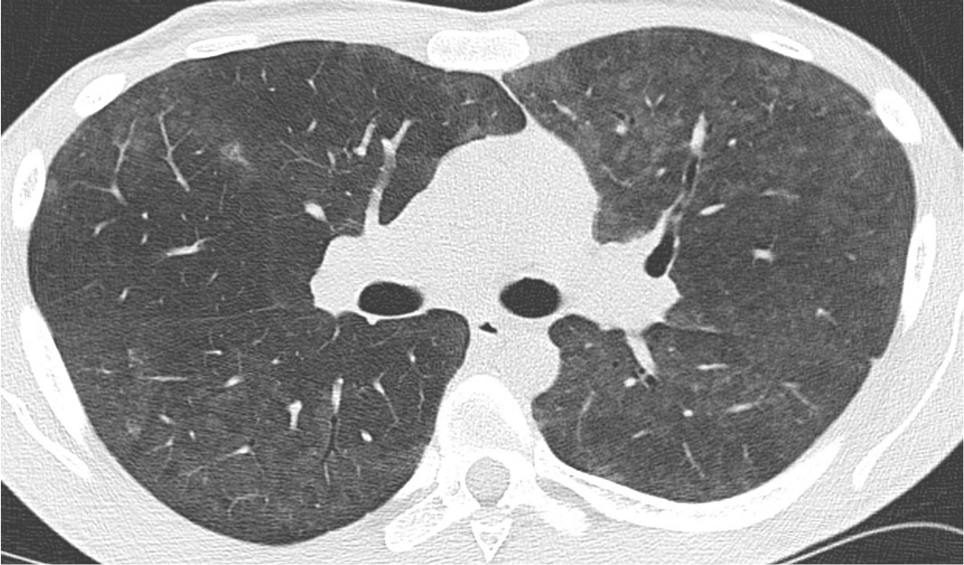
A man in his late 20s with HIV (CD4+ count, 8 cells/μ L) not on ART presented with shortness of breath. Axial CT image of the chest in lung windows demonstrated diffuse, ill-defined centrilobular ground-glass nodules throughout the lungs (Figure 38). Pneumocystis DFA staining and PCR test on a sputum sample were positive.26–28
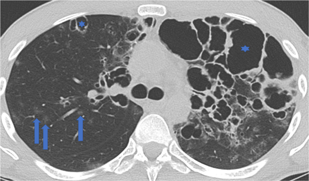
A man in his late 20s with advanced HIV not on ART presented with persistent dyspnea, hypoxemia, tachypnea, and fever. Axial CT image of the chest in lung windows demonstrated scattered ground-glass nodules (Figure 39, arrows) and numerous superimposed cavitary lesions (Figure 39, asterisks). Pneumocystis DFA staining from a bronchoalveolar lavage specimen was positive.26–28
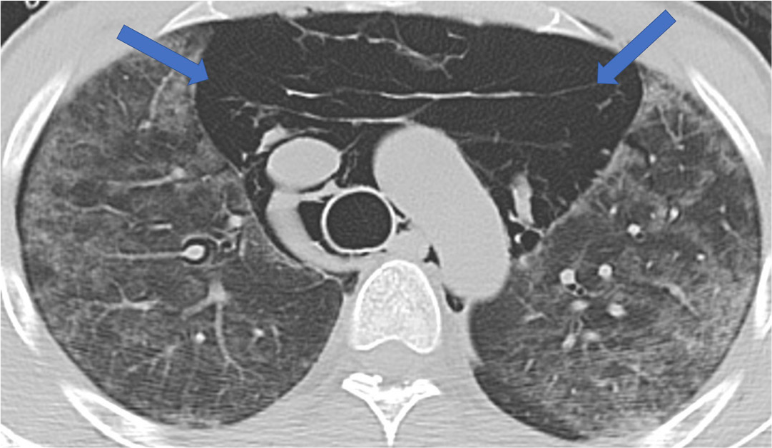
A man in his mid-20s with recently diagnosed HIV (CD4+ count, 12 cells/μL) not on ART presented with progressive shortness of breath, dry cough, pleuritic chest pain, fevers, and lightheadedness. Initial CT imaging of the chest (not pictured) demonstrated ground-glass opacities throughout the lungs. He underwent bronchoscopy with bronchoalveolar lavage specimen positive for Pneumocystis jirovecii by DFA staining. He initiated treatment with trimethoprim-sulfamethoxazole (TMP-SMX). Six days after initiation of therapy, he experienced acute respiratory decompensation. Repeat CT imaging demonstrated patchy ground-glass opacities as well as extensive pneumomediastinum (Figure 40, arrows).26–28
Progressive Mu.pngocal Leukoencephalopathy
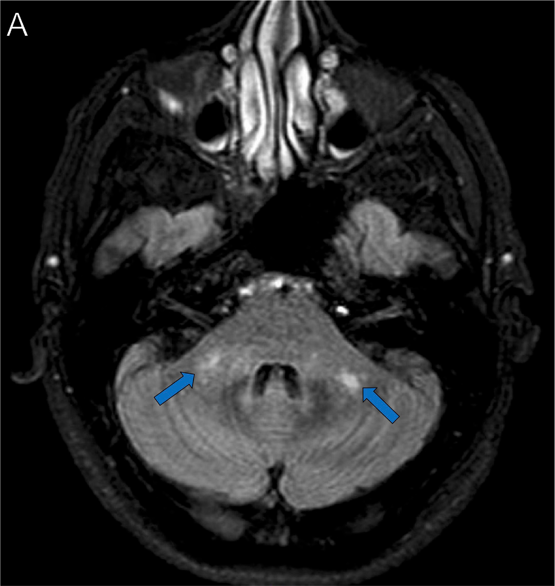
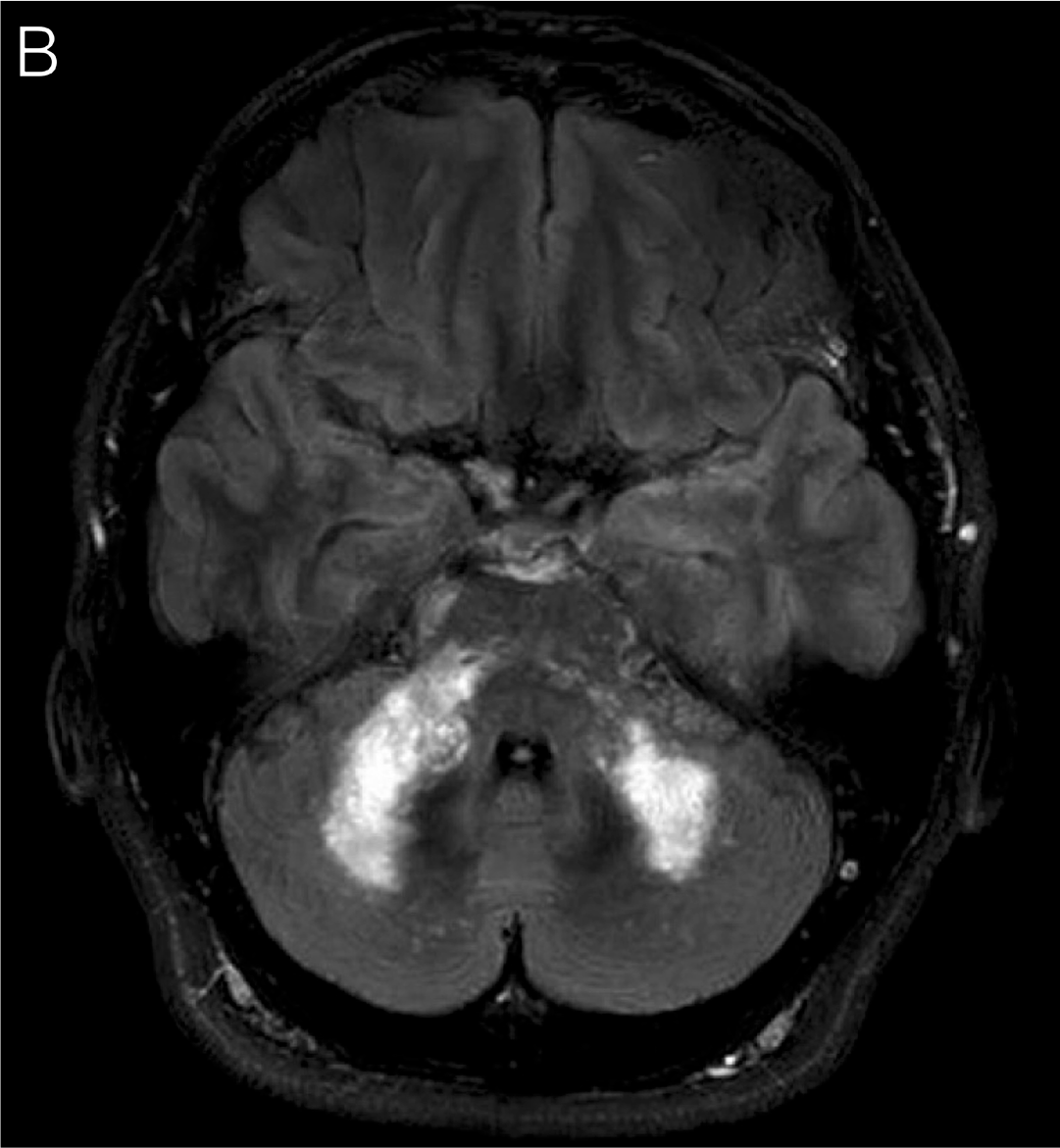
A man in his mid-40s with recently diagnosed HIV (CD4+ count, 61 cells/μL) not on ART presented while asymptomatic for enrollment on study protocol. Brain MRI showed multiple foci of signal abnormality within the middle cerebellar peduncles and bilateral cerebellum that demonstrated mild restricted diffusion and T1 hypointensity (Figure 41A, arrows), consistent with progressive mu.pngocal leukoencephalopathy (PML). Two months after ART initiation, he developed ataxia as well as facial and lower-extremity numbness. Repeat brain MRI showed substantial progression of white matter changes (Figure 41B), concerning for PML IRIS.29,30
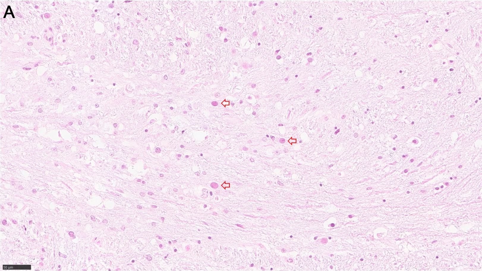
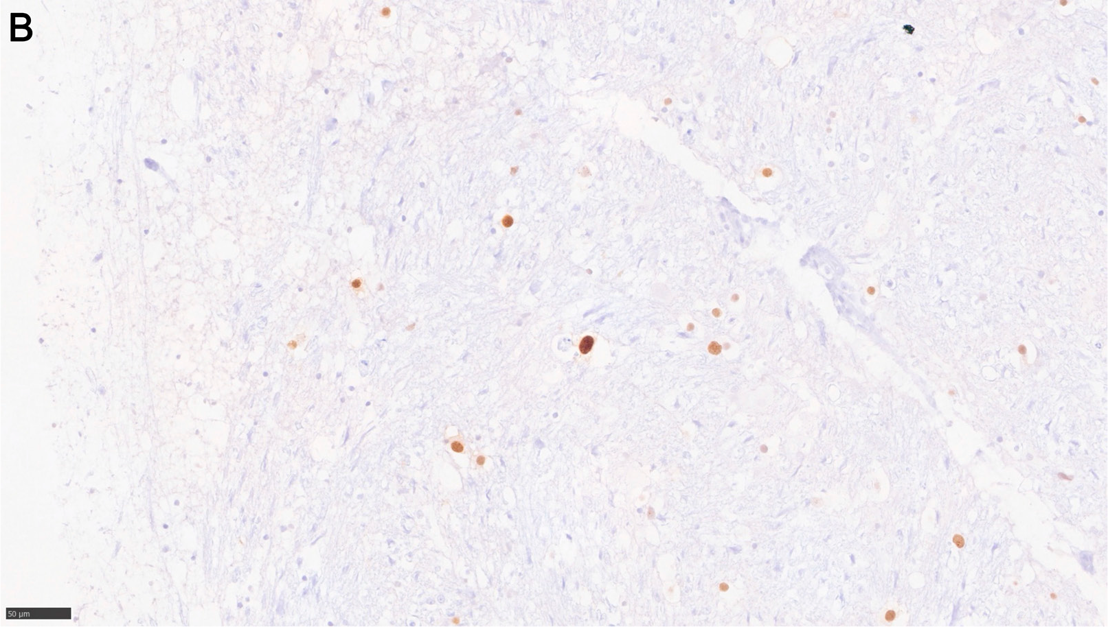
A man in his early 30s presented to the hospital with 2 weeks of progressive aphasia, new-onset memory loss, and right-sided weakness. An HIV test was positive, and his CD4+ count was 4 cells/µL. Brain MRI showed increased T2/FLAIR and decreased T1 signal involving the white matter of the left frontal occipitoparietal and posteromedial temporal lobes, characteristics suggestive of PML. CSF was positive for human polyomavirus 2 (John Cunningham [JC] virus). The patient was started on dexamethasone and ART, but he developed progressively worsening symptoms, altered mental status, and ultimately was found without pulse. Resuscitative efforts were unsuccessful. Brain autopsy specimens showed viral cytopathic changes (Figure 42A, arrows) and positive simian virus 40 (SV40) immunohistochemistry (Figure 42B), consistent with JC virus infection.29,30
Strongyloidiasis
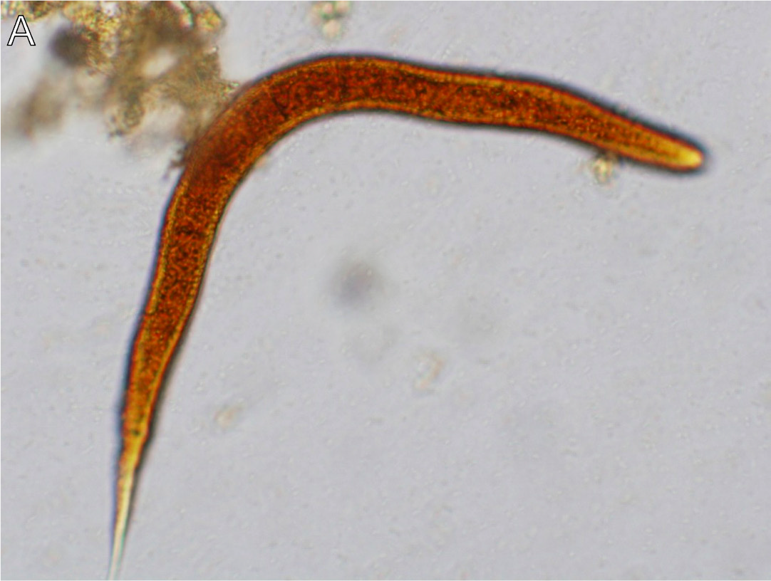
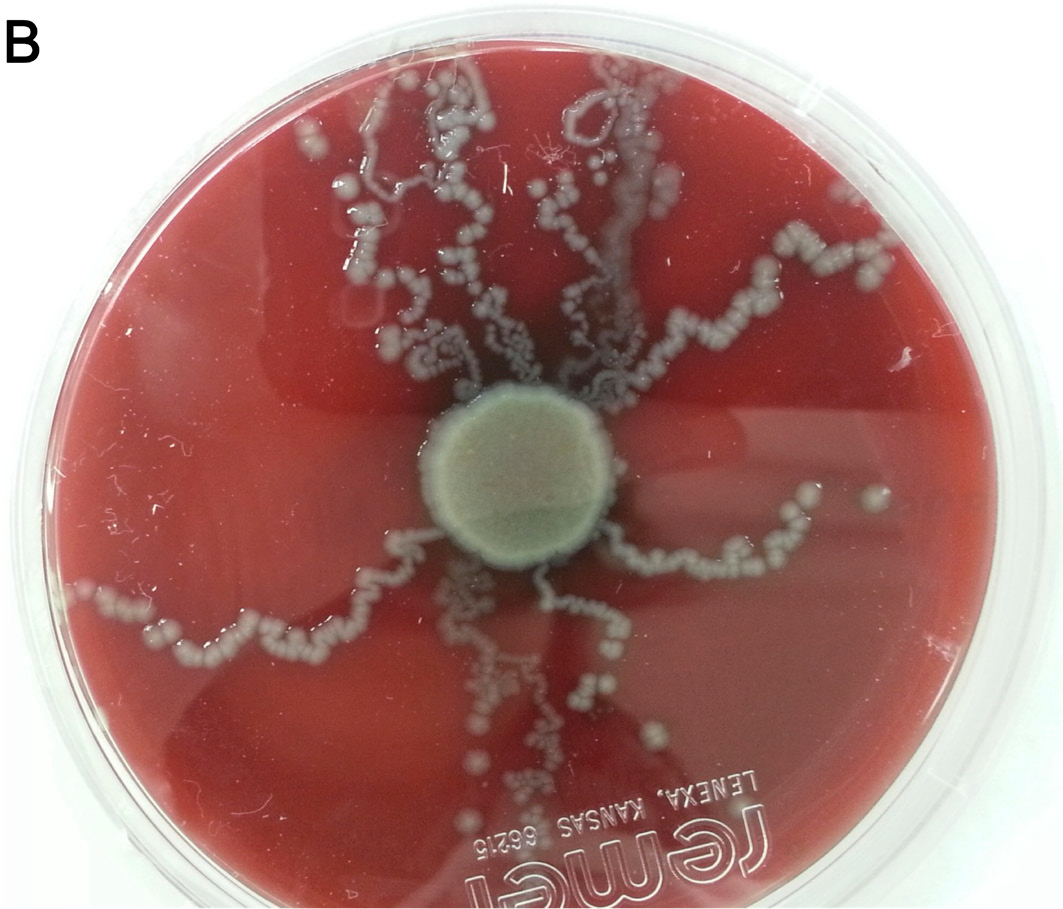
Aman in his late 30s originally from El Salvador (who had been in the US for >10 years) presented with diarrhea for the preceding 3 months. He was diagnosed with HIV and had a CD4+ count of 43 cells/μL. Work-up for the diarrhea included a stool ova and parasite exam, which revealed rhabd.pngorm larvae of Strongyloides stercoralis (Figure 43A). Stool culture revealed visible tracks of bacterial colonies along the path of larvae migration (Figure 43B).31 He was treated with oral ivermectin for 2 days with quantitative decline in larval forms, but he continued to have diarrhea. He reinitiated treatment for hyperinfection syndrome with ivermectin for an additional 10 days, with subsequent clearance of stool ova and parasite examinations and improvement in his diarrhea.
Toxoplasmosis
Aman in his early 30s originally from Nicaragua with no prior medical history presented with seizures, aphasia, and right-sided weakness. He was found to have numerous ring-enhancing lesions, including an irregularly shaped lesion within the left parietal lobe measuring 2.3 cm with surrounding edema (Figures 44A-B, arrows). An HIV test was positive and his CD4+ count was 59 cells/µL. Biopsy of the parietal lesion revealed tachyzoites and tissue cysts filled with bradyzoites on H&E (Figure 44C), with immunostaining showing tachyzoites (Figure 44D), and GMS stain highlighting tissue cysts (Figure 44E). He was treated with TMP-SMX, on which he improved.32
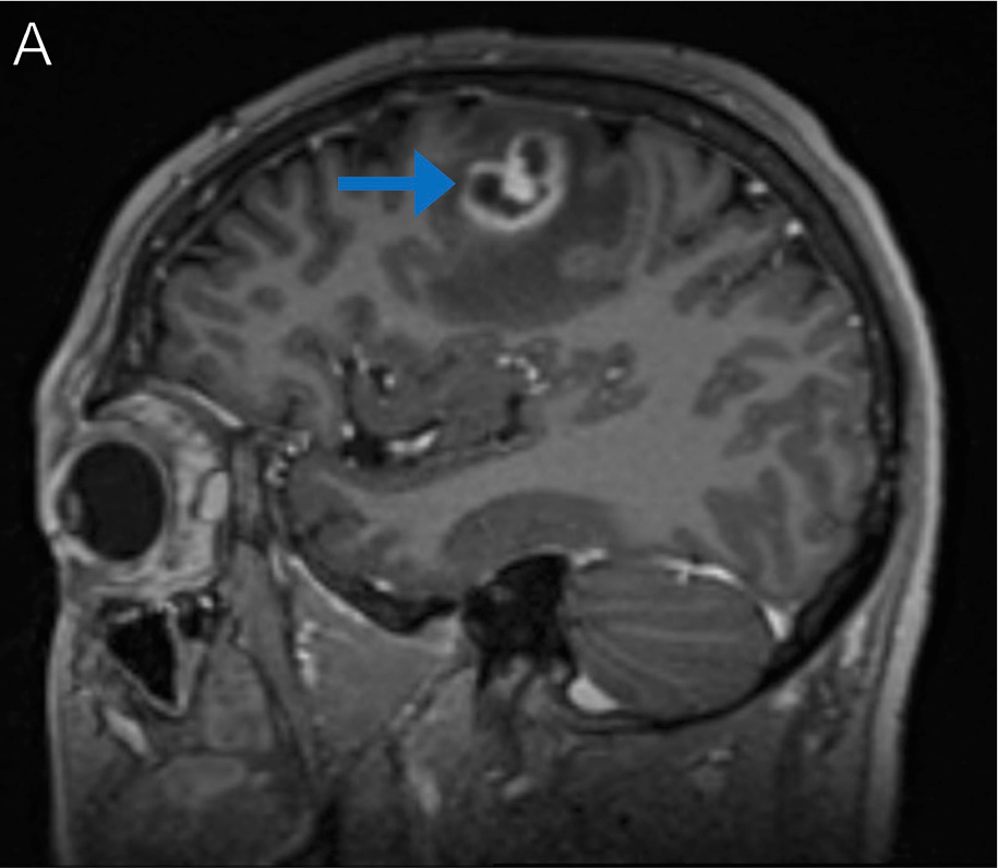
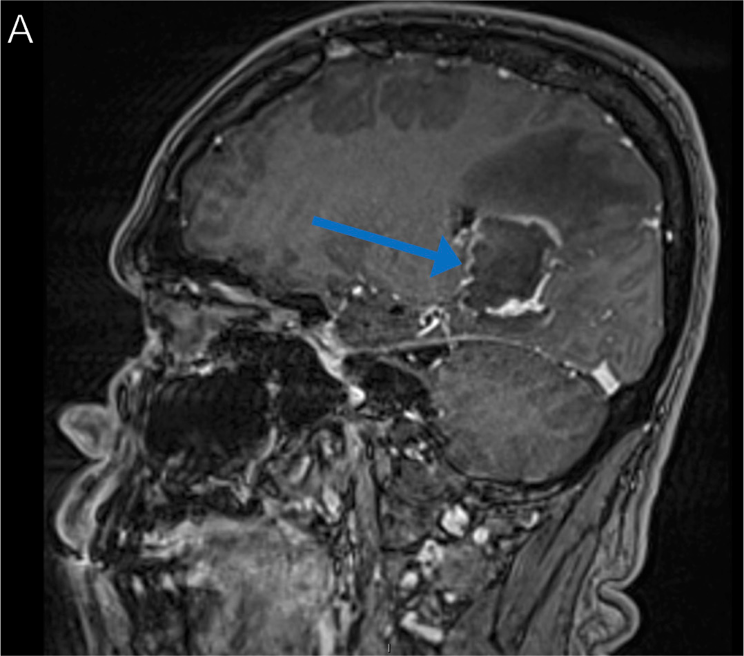
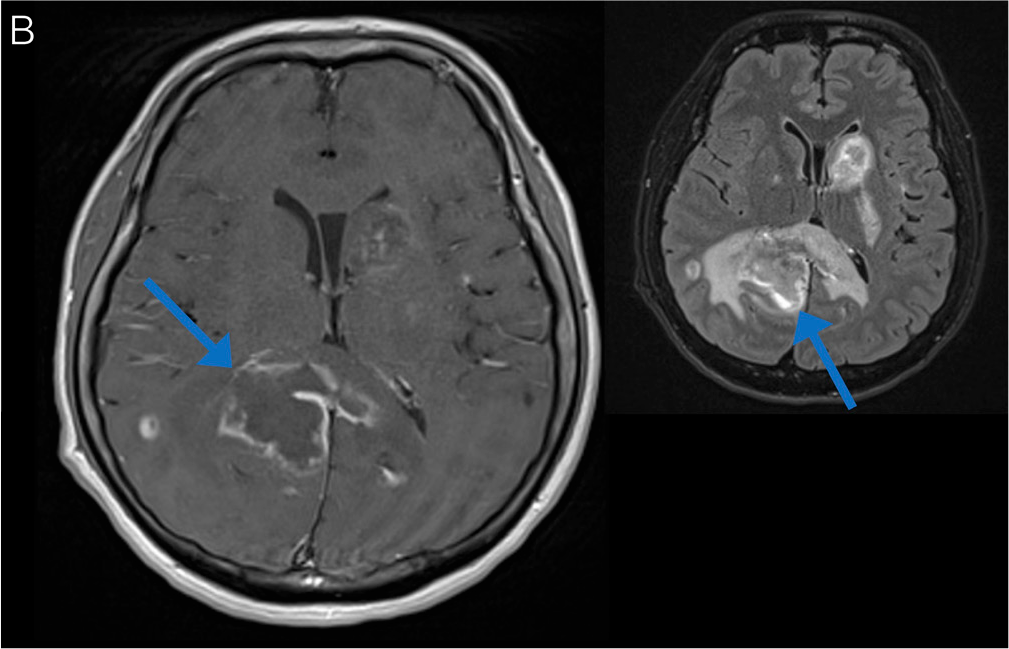
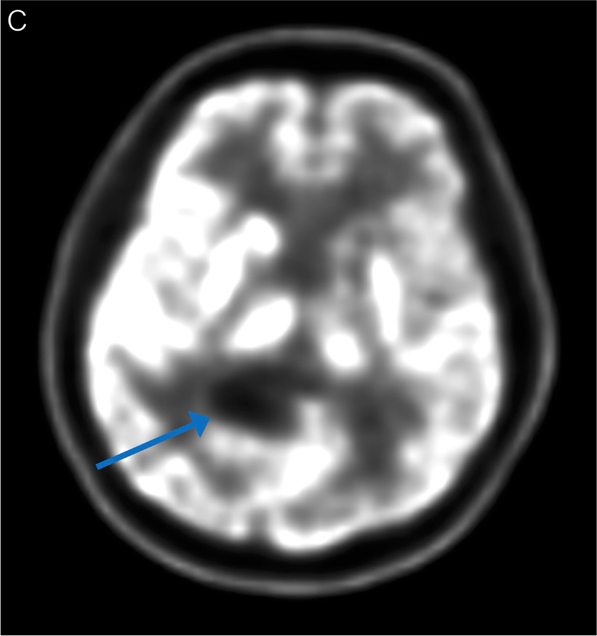
A man in his late 40s presented with progressively worsening headache, confusion, memory loss, and fatigue. An HIV test was positive. MRI of the brain revealed a dominant 3 × 3 × 5-cm irregular rim-enhancing mass with central restricted diffusion and extensive edema in the splenium callosum and right occipital lobe, causing local mass effect, compression of the posterior lateral ventricle, and dilation of the right temporal horn (Figures 45A-B, arrows). Toxoplasma PCR test from CSF was positive. He started TMP-SMX and dexamethasone (for 2 weeks); however, a repeat brain MRI did not demonstrate substantial improvement. A PET-CT scan of the brain was subsequently completed 4 weeks after treatment initiation, and did not demonstrate increased uptake in the occipital lesion (Figure 45C, arrow), more consistent with toxoplasmosis as opposed to primary central nervous system (CNS) lymphoma.33 He continued treatment with TMP-SMX and initiated ART, although he developed a rash a few weeks later that resolved after switching TMP-SMX to clindamycin and pyrimethamine, on which he improved, with decreasing size in the CNS lesions.32
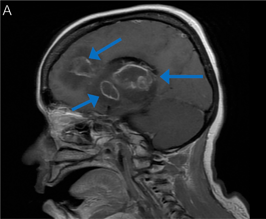
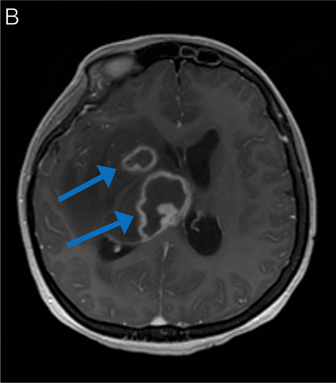
A woman in her late 30s originally from El Salvador presented with headaches, diplopia, word-finding problems, and left-sided weakness. An HIV test was positive. MRI of the brain showed large necrotic lesions with weak peripheral enhancement centered in the right basal ganglia and the anterior body and genu of the corpus callosum (Figures 46A-B, arrows). A serum Toxoplasma IgG test was positive. She underwent brain biopsy, with pathology demonstrating cysts and tachyzoites, consistent with toxoplasmosis. She began treatment with TMP-SMX, on which her motor symptoms improved.32
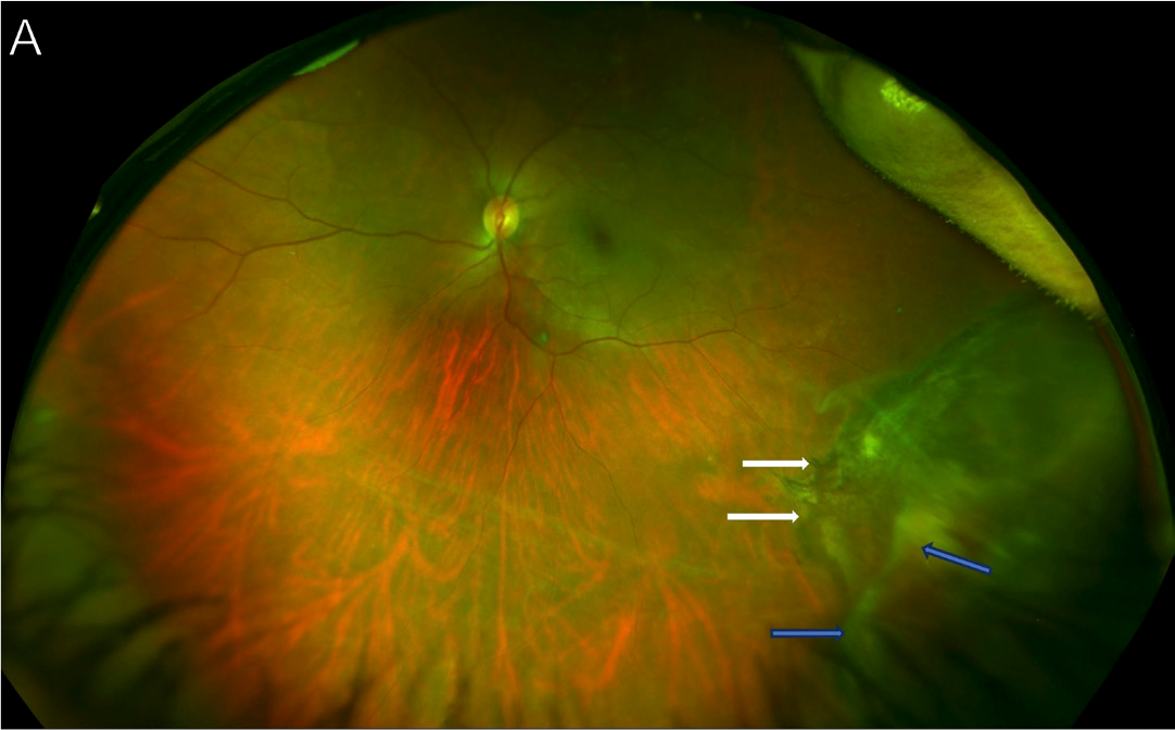
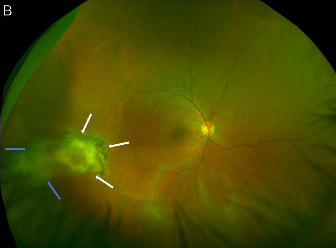
A man in his late 30s with HIV (CD4+ count, 42 cells/μL)
not on ART presented with bilateral blurry vision for the preceding 4 weeks. Fundus photos of left eye showed a peripheral inferior chorioretinal lesion with pigment hypertrophy (Figure 47A, white arrows) and overlying vitritis (Figure 47A, blue arrows). His right eye also showed a temporal chorioretinal lesion (Figure 47B, white arrows) and vitritis (Figure 47B, blue arrows) consistent with bilateral toxoplasma chorioretinitis. An aqueous fluid PCR test was positive for Toxoplasma gondii.32
Varicella-Zoster Virus
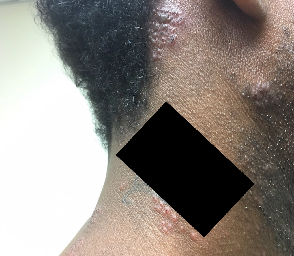
A man in his mid-40s with advanced HIV and presumed disseminated MAC (pancytopenia and poorly formed granulomas on bone marrow biopsy) on treatment for about 1 year presented with pruritic rash on the right side of his neck (Figure 48) and right ear pain for the preceding 4 days. Examination showed vesicles clustered on an erythematous base along the right C2-C3 dermatome and over the right posterior ear and right earlobe. There were no lesions on the tympanic membrane. Varicella-zoster virus (VZV) PCR testing on a swab of a representative skin lesion was positive. He was treated with valacyclovir for 10 days and had resolution of the rash.34
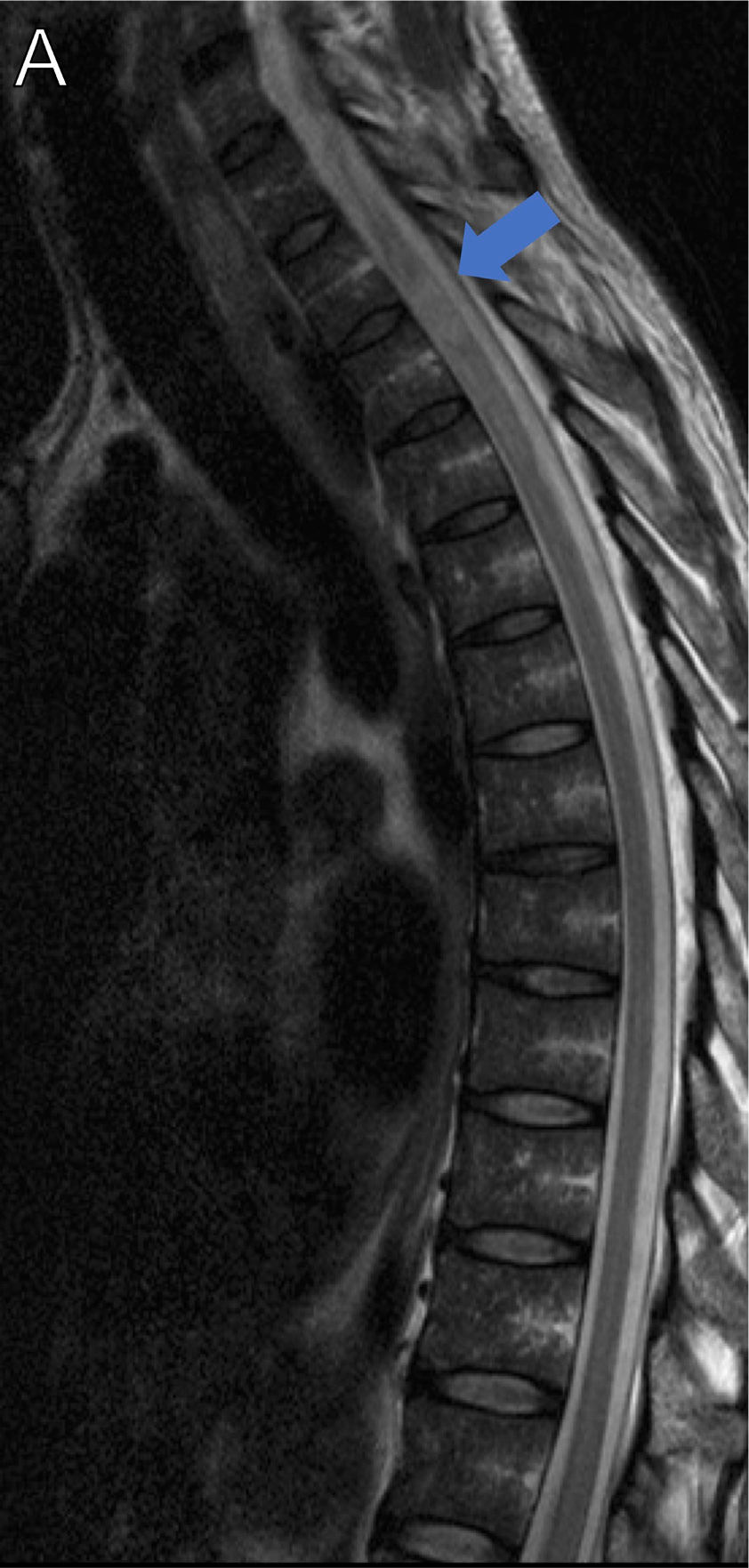
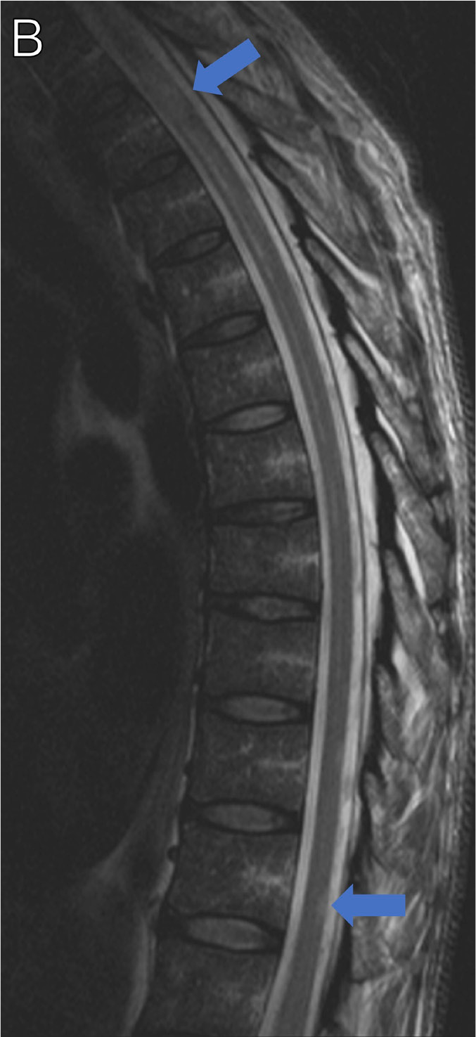
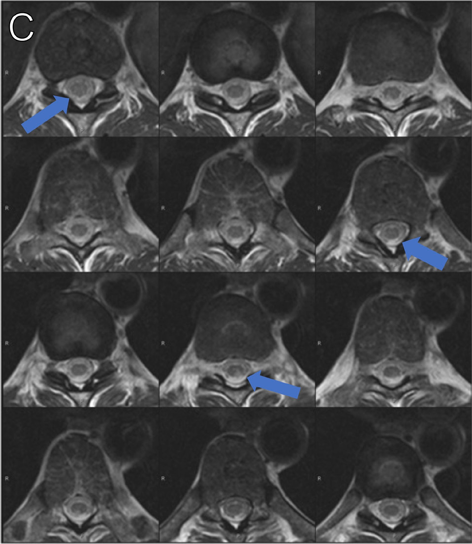
A man in his 50s presented with a 4-day history of leftsided chest pain and progressive left leg weakness. He had a 3-day history of a vesicular rash at the nipple line that wrapped around posteriorly, staying on the left side of midline. His exam was notable for left lower-extremity weakness and decreased sensation to pinprick and temperature of his right lower extremity. Imaging revealed spinal cord abnormalities at C7 and T3-T4, with widening and enhancement of the cord at T3 (Figure 49A, arrow). He underwent a lumbar puncture that revealed lymphocytic pleocytosis, an elevated total protein level, and PCR sequencing was positive for VZV. The presentation was thought to be consistent with transverse myelitis with Brown-Séquard syndrome secondary to VZV. The patient was initiated on IV acyclovir and corticosteroids with initial improvement, but then had subsequent worsening with new hyperintense lesions in the spinal cord (Figures 49B-C, arrows). He continued on IV acyclovir with slow improvement.34
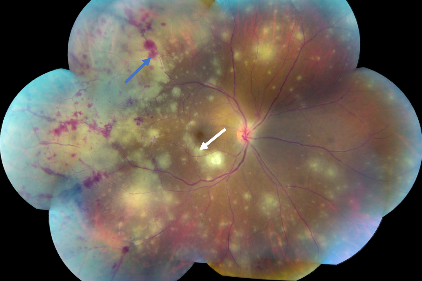
A woman in her early 40s with HIV (CD4+ count, 14 cells/μL) not on ART presented with acute peripheral vision loss in her right eye. Aqueous humor PCR test was positive for VZV.34 Dilated eye exam showed circumferential areas of patchy retinitis with confluence of the lesions from 7:00 to 12:00, as well as retinitis in the posterior pole including the macula (Figure 50, white arrow). Associated features include diffuse vasculitis and retinal hemorrhages (Figure 50, blue arrow). There was notable absence of substantial vitritis.
Acknowledgments
All patients described were evaluated at the National Institutes of Health Clinical Center and had been enrolled as clinical study participants. They had provided written informed consent and were enrolled on the clinical protocols NCT00286767 or NCT02147405. These protocols were approved by the National Institutes of Health Institutional Review Board. Funding for this study was provided in part by the Division of Intramural Research program of the National Institute of Allergy and Infectious Diseases.
Disclaimer
This research was supported by the Intramural Research Program of the National Institutes of Health (NIH). The contributions of the NIH authors are considered Works of the United States Government. The findings and conclusions presented in this paper are those of the authors and do not necessarily reflect the views of the NIH or the US Department of Health and Human Services.
The IAS-USA ide.pngied and mitigated ahead of time any possible conflicts of interest that may influence CME activities with regard to exposition or conclusion. All financial relationships with ineligible companies for the contributors, planners, and reviewers are below.
Financial relationships with ineligible companies within the past 24 months: Dr Bellur reported receiving payment as an employee of Sanofi Pharmaceuticals. (Updated August 10, 2025) Dr Dampier reported no relevant financial relationships with ineligible companies. (Updated April 3, 2025) Dr Epling reported no relevant financial relationships with ineligible companies. (Updated April 3, 2025) Ms Laidlaw reported no relevant financial relationships with ineligible companies. (Updated April 3, 2025) Dr Magone De Quadros Costa reported no relevant financial relationships with ineligible companies. (Updated April 3, 2025) Dr Manion reported no relevant financial relationships with ineligible companies. (Updated April 4, 2025) Dr Sereti reported no relevant financial relationships with ineligible companies. (Updated April 3, 2025) Dr Sheikh reported no relevant financial relationships with ineligible companies. (Updated August 10, 2025) Dr Sirajuddin reported no relevant financial relationships with ineligible companies. (Updated August 28, 2025)
Reviewer 1 reported consulting or advisor fees from Generate Biomedicines and Gilead Sciences, Inc. (Updated July 23, 2025) Reviewer 2 reported no relevant financial relationships with ineligible companies. (Updated August 7, 2025) Reviewer 3 reported no relevant financial relationships with ineligible companies. (Updated August 7, 2025)
References
R Rajasingham, NP Govender, A Jordan, et al. Theglobalburden of HIV-associated cryptococcal infection in adults in 2020: a modelling analysis. Lancet Infect Dis. 2022; 22(12): 1748–1755. doi: 10.1016/S1473-3099(22)00499-6.
N Ford, P Ehrenkranz, J Jarvis. Advanced HIV as a neglected disease. N Engl J Med. 2024; 390(6): 487–489. doi: 10.1056/NEJMp2313777.
Centers for Disease Control and Prevention. Monitoring selected national HIV prevention and care objectives by using HIV surveillance data United States and 6 territories and freely associated states, 2022. Published May 21, 2024. Accessed August 27, 2025. https://stacks.cdc.gov/view/cdc/156511.
C Bielick, A Strumpf, S Ghosal, T McMurry, KA McManus. National hospitalization rates and in-hospital mortality rates of HIV-related opportunistic infections in the United States, 2011-2018. Clin Infect Dis. 2024; 79(2): 487–497. doi: 10.1093/cid/ciae051.
G Meintjes, G Maartens. HIV-associated tuberculosis. N Engl J Med. 2024; 391(4): 343–355. doi: 10.1056/NEJMra2308181.
RK Gupta, SB Lucas, KL Fielding, SD Lawn. Prevalence of tuberculosis in post-mortem studies of HIV-infected adults and children in resource-limited settings: a systematic review and meta-analysis. AIDS. 2015; 29(15): 1987–2002. doi: 10.1097/QAD.0000000000000802.
Panel on Guidelines for the Prevention and Treatment of Opportunistic Infections in Adults and Adolescents With HIV. Guidelines for the prevention and treatment of opportunistic infections in adults and adolescents with HIV: bartonellosis. National Institutes of Health, Centers for Disease Control and Prevention, HIV Medicine Association, and Infectious Diseases Society of America. 2023. Updated November 14, 2023. Accessed August 27, 2025. https://clinicalinfo.hiv.gov/en/guidelines/hiv-clinical-guidelines-adult-and-adolescent-opportunistic-infections/bartonellosis.
Panel on Guidelines for the Prevention and Treatment of Opportunistic Infections in Adults and Adolescents With HIV. Guidelines for the prevention and treatment of opportunistic infections in adults and adolescents with HIV: candidiasis (mucocutaneous). National Institutes of Health, Centers for Disease Control and Prevention, HIV Medicine Association, and Infectious Diseases Society of America. 2023. Updated September 16, 2024. Accessed August 27, 2025. https://clinicalinfo.hiv.gov/en/guidelines/hiv-clinical-guidelines-adult-and-adolescent-opportunistic-infections/candidiasis.
Panel on Guidelines for the Prevention and Treatment of Opportunistic Infections in Adults and Adolescents With HIV. Guidelines for the prevention and treatment of opportunistic infections in adults and adolescents with HIV: cryptococcosis. National Institutes of Health, Centers for Disease Control and Prevention, HIV Medicine Association, and Infectious Diseases Society of America. 2024. Updated October 29, 2024. Accessed August 27, 2025. https://clinicalinfo.hiv.gov/en/guidelines/hiv-clinical-guidelines-adult-and-adolescent-opportunistic-infections/cryptococcosis.
M Manion, I Sereti. A 34-year-old man with HIV/AIDS and a cavitary pulmonary lesion. Chest. 2021; 160(1): e35–e38. doi: 10.1016/j.chest.2021.01.086.
Panel on Guidelines for the Prevention and Treatment of Opportunistic Infections in Adults and Adolescents With HIV. Guidelines for the prevention and treatment of opportunistic infections in adults and adolescents with HIV: cytomegalovirus diease. National Institutes of Health, Centers for Disease Control and Prevention, HIV Medicine Association, and Infectious Diseases Society of America. 2021. Updated July 01, 2021. Accessed August 27, 2025. https://clinicalinfo.hiv.gov/en/guidelines/hiv-clinical-guidelines-adult-and-adolescent-opportunistic-infections/cytomegalovirus.
KF Faldetta, S Kattakuzhy, HW Wang, I Sereti, V Sheikh. Cytomegalovirus immune reconstitution inflammatory syndrome manifesting as acute appendicitis in an HIV-infected patient. BMC Infect Dis. 2014; 14: 313. doi: 10.1186/1471-2334-14-313.
V Sheikh, M Caplan, EM Wilson, et al. Cytomegalovirus immune reconstitution inflammatory syndrome manifesting as sialadenitis in an HIV-infected patient. AIDS. 2013; 27(11): 1833–1835. doi: 10.1097/QAD.0b013e328362d8b0.
M Huguet, JT Navarro, J Molto, JM Ribera, G Tapia. Diffuse large B-cell lymphoma in the HIV setting. Cancers (Basel). 2023; 15(12): 3191. doi: 10.3390/cancers15123191.
Panel on Guidelines for the Prevention and Treatment of Opportunistic Infections in Adults and Adolescents With HIV. Guidelines for the prevention and treatment of opportunistic infections in adults and adolescents with HIV: histoplasmosis. National Institutes of Health, Centers for Disease Control and Prevention, HIV Medicine Association, and Infectious Diseases Society of America. 2024. Updated October 29, 2024. Accessed August 27, 2025. https://clinicalinfo.hiv.gov/en/guidelines/hiv-clinical-guidelines-adult-and-adolescent-opportunistic-infections/histoplas-mosis.
M Manion, A Boulougoura, N Naqvi, et al. Polyfunctional antigen specific CD4+ T cell responses in patients with human immunodeficiency virus/AIDS and histoplasmosis immune reconstitution inflammatory syndrome. Clin Infect Dis. 2023; 76(3): 531–534. doi: 10.1093/cid/ciac514.
KA Lurain, R Ramaswami, LT Krug, et al. HIV-associated cancers and lymphoproliferative disorders caused by Kaposi sarcoma herpesvirus and Epstein-Barr virus. Clin Microbiol Rev. 2024; 37(3): e0002223. doi: 10.1128/cmr.00022-23.
Panel on Guidelines for the Prevention and Treatment of Opportunistic Infections in Adults and Adolescents With HIV. Guidelines for the prevention and treatment of opportunistic infections in adults and adolescents with HIV: human herpesvirus-8 disease. National Institutes of Health, Centers for Disease Control and Prevention, HIV Medicine Association, and Infectious Diseases Society of America. 2024. Updated April 23, 2025. Accessed August 27, 2025. https://clinicalinfo.hiv.gov/en/guidelines/hiv-clinical-guidelines-adult-and-adolescent-opportunistic-infections/human-herpesvirus.
Panel on Guidelines for the Prevention and Treatment of Opportunistic Infections in Adults and Adolescents With HIV. Guidelines for the prevention and treatment of opportunistic infections in adults and adolescents with HIV: human papillomavirus disease. National Institutes of Health, Centers for Disease Control and Prevention, HIV Medicine Association, and Infectious Diseases Society of America. 2024. Updated July 9, 2024. Accessed August 27, 2025. https://clinicalinfo.hiv.gov/en/guidelines/hiv-clinical-guidelines-adult-and-adolescent-opportunistic-infections/human.
Panel on Guidelines for the Prevention and Treatment of Opportunistic Infections in Adults and Adolescents With HIV. Guidelines for the prevention and treatment of opportunistic infections in adults and adolescents with HIV: disseminated mycobacterium avium complex disease. National Institutes of Health, Centers for Disease Control and Prevention, HIV Medicine Association, and Infectious Diseases Society of America. 2024. Updated August 15, 2024. Accessed August 27, 2025. https://clinicalinfo.hiv.gov/en/guidelines/hiv-clinical-guidelines-adult-and-adolescent-opportunistic-infections/disseminated.
BP Epling, M Manion, E Laidlaw, et al. When location and timing are at the heart of the diagnosis. Clin Infect Dis. 2024; 79(3): 775–777. doi: 10.1093/cid/ciae179.
Panel on Guidelines for the Prevention and Treatment of Opportunistic Infections in Adults and Adolescents With HIV. Guidelines for the prevention and treatment of opportunistic infections in adults and adolescents with HIV: mycobacterium tuberculosis. National Institutes of Health, Centers for Disease Control and Prevention, HIV Medicine Association, and Infectious Diseases Society of America. 2024. Updated May 2, 2024. Accessed August 27, 2025. https://clinicalinfo.hiv.gov/en/guidelines/hiv-clinical-guidelines-adult-and-adolescent-opportunistic-infections/mycobacterium.
DC Hsu, KF Faldetta, L Pei, et al. A paradoxical treatment for a paradoxical condition: infliximab use in three cases of mycobacterial IRIS. Clin Infect Dis. 2016; 62(2): 258–261. doi: 10.1093/cid/civ841.
M Manion, N Lynn, L Pei, et al. To induce immune reconstitution inflammatory syndrome or suppress it: the spectrum of Mycobacterium genavense in the antiretroviral era. Clin Infect Dis. 2021; 72(2): 315–318. doi: 10.1093/cid/civ841.
M Manion, CA Dulanto, L Pei, et al. Disseminated Mycobacterium marinum in human immunodeficiency virus unmasked by immune reconstitution inflammatory syndrome. J Infect Dis. 2021; 224(3): 453–457. doi: 10.1093/infdis/jiaa769.
Panel on Guidelines for the Prevention and Treatment of Opportunistic Infections in Adults and Adolescents With HIV. Guidelines for the prevention and treatment of opportunistic infections in adults and adolescents with HIV: pneumocystis pneumonia. National Institutes of Health, Centers for Disease Control and Prevention, HIV Medicine Association, and Infectious Diseases Society of America. 2024. Updated September 16, 2024. Accessed August 27, 2025. https://clinicalinfo.hiv.gov/en/guidelines/hiv-clinical-guidelines-adult-and-adolescent-opportunistic-infections/pneumocystis.
BP Epling, M Manion, A Sirajuddin, et al. Long-term outcomes of patients with HIV and Pneumocystis jirovecii pneumonia in the antiretroviral therapy era. Open Forum Infect Dis. 2023; 10(8): ofad408. doi: 10.1093/ofid/ofad408.
EG McDonald, A Afshar, B Assiri, et al. Pneumocystis jirovecii pneumonia in people living with HIV: a review. Clin Microbiol Rev. 2024; 37(1): e0010122. doi: 10.1128/cmr.00101-22.
I Cortese, DS Reich, A Nath. Progressive mu.pngocal leukoencephalopathy and the spectrum of JC virus-related disease. Nat Rev Neurol. 2021; 17(1): 37–51. doi: 10.1038/s41582-020-00427-y.
Panel on Guidelines for the Prevention and Treatment of Opportunistic Infections in Adults and Adolescents With HIV. Guidelines for the prevention and treatment of opportunistic infections in adults and adolescents with HIV: progressive mu.pngocal leukoencephalopathy/JC virus infection. National Institutes of Health, Centers for Disease Control and Prevention, HIV Medicine Association, and Infectious Diseases Society of America. 2022. Updated October 19, 2025. Accessed August 27, 2025. https://clinicalinfo.hiv.gov/en/guidelines/hiv-clinical-guidelines-adult-and-adolescent-opportunistic-infections/progressive.
CA Gordon, J Utzinger, S Muhi, et al. Strongyloidiasis. Nat Rev Dis Primers. 2024; 10(1): 6. doi: 10.1038/s41572-023-00490-x.
Panel on Guidelines for the Prevention and Treatment of Opportunistic Infections in Adults and Adolescents With HIV. Guidelines for the prevention and treatment of opportunistic infections in adults and adolescents with HIV: toxoplasmosis. National Institutes of Health, Centers for Disease Control and Prevention, HIV Medicine Association, and Infectious Diseases Society of America. 2025. Updated September 16, 2024. Accessed August 27, 2025. https://clinicalinfo.hiv.gov/en/guidelines/hiv-clinical-guidelines-adult-and-adolescent-opportunistic-infections/toxoplasmosis.
MA Pierce, MD Johnson, RJ Maciunas, et al. Evaluating contrast-enhancing brain lesions in patients with AIDS by using positron emission tomography. Ann Intern Med. 1995; 123(8): 594–598. doi: 10.7326/0003-4819-123-8-199510150-00005.
Panel on Guidelines for the Prevention and Treatment of Opportunistic Infections in Adults and Adolescents With HIV. Guidelines for the prevention and treatment of opportunistic infections in adults and adolescents with HIV: varicella-zoster virus disease. National Institutes of Health, Centers for Disease Control and Prevention, HIV Medicine Association, and Infectious Diseases Society of America. 2022. Updated September 7, 2022. Accessed August 27, 2025. https://clinicalinfo.hiv.gov/en/guidelines/hiv-clinical-guidelines-adult-and-adolescent-opportunistic-infections/varicella-zoster.